


This is a joint FSA and FSS publication.

1. Introduction
The Food Standards Agency (FSA) and Food Standards Scotland (FSS) have undertaken a safety assessment for application regarding the use of glycolipids as a new food additive, submitted by the company, LANXESS Deutschland GmbH under Regulation (EC) No 1331/2008.
The glycolipids compound is produced by the fermentation of the edible mushroom Dacryopinax spathularia, strain MUCL 53181 (internal identifier: FU50088). The product undergoes microfiltration to remove fungal cells, is purified, then pasteurised and spray-dried.
The resulting substance, glycolipids, has antimicrobial effects and, thus, is proposed for use as a preservative in beverages.
2. Assessment
2.1. Overview
As part of the Regulated Products authorisation process, the Joint Expert Group on Additives, Enzymes, and other Regulated Products (AEJEG) were requested by the FSA and FSS to provide advice on the application for the safety of the Application of glycolipids (AM-1) for use as a new food additive under Regulation (EC) No 1331/2008.
The glycolipids compound is produced by the fermentation of the edible mushroom Dacryopinax spathularia, strain MUCL 53181 (internal identifier: FU50088).
Satisfactory information regarding proposed specifications, details of the manufacturing process, presence of impurities, stability of the additive and fate in foods, dietary exposure assessment, and toxicological data, were submitted.
The AEJEG concluded that no concern was raised from the manufacturing process. The AEJEG considered that the toxicological information provided by the applicant that formed the basis of the risk assessment was satisfactory.
The AEJEG raised concerns, which were subsequently endorsed by both the COT and ACMSF, that a chronic consumption of beverages containing the AM-1 additive could lead to the ingestion of a small dose of a product which retains antimicrobial properties even after deacylation.
To address these concerns, a study on the faecal and caecal matter of SD rats, treated by oral gavage up to 1000mg/kg/bw/day over 90 days was then provided. This was reviewed by the FSA/FSS which concluded that the results do not indicate any biologically meaningful changes in microbiome composition at doses as high as 1000mg/kg bw/day following 90 days of exposure.
Overall, the FSA and FSS concluded there was no toxicity, genotoxicity or gut microbiome concerns for glycolipids produced using the strain MUCL 53181 and agreed on a NOAEL of 1000 mg/kg bw per day based on the 90-day rat study, and the use of a total uncertainty factor of 300 to give an ADI of 3.3 mg/kg bw per day.
2.2. Identity and characterisation of the product
The Applicant has proposed the use of glycolipids from Dacryopinax spathularia in accordance with the EFSA guidance for submission of food additives (EFSA, 2012) this food additive belongs to the category of “Additives derived from botanical sources”.
The Applicant proposed the following names for the food additive:
-
Glycolipids
-
AM-1
Throughout the application the applicant provided an alternative name for the glycolipid product, as AM-1, this is used as an identifier for the glycolipids to ensure consistency with the EFSA’s Scientific Opinion adopted on 4 May 2021 (EFSA, 2021). In the European Union, Switzerland, Norway, Liechtenstein, New Zealand, Australia, Mexico, the United States and other jurisdiction, AM-1 is marketed under the specific trademark name Nagardo©. For clarity, the term AM-1 will be used to refer to glycolipids from this point forwards.
AM-1 is produced via fermentation of the edible mushroom Dacryopinax spathularia, strain MUCL 53181 (internal identifier: FU50088). This was collected in French Guiana, an Overseas Department of the French Republic, and obtained before 12 October 2014. The applicant stated that the strain MUCL 53181 has optimum performance and yield in the fermentation process. Only this one specific strain is used to produce the AM-1; and this strain is the property of IMD Natural Solutions GmbH, who are part of the Lanxess company.
The AEJEG Members highlighted that the safety assessment and its conclusions should apply to glycolipids produced using specifically the strain MUCL 53181.
-
Name: Glycolipids
-
Source: Dacryopinax spathularia strain MUCL 53181
-
E number: E 246
-
Synonyms: AM-1
-
Chemical name: Long-chain glycolipid congeners
-
CAS number: 2205009-17-0
-
Solubility: 10 g/L in water
-
Molecular formula: variable, depending on the acyl groups
-
Molecular weight: variable, depending on the acyl groups
The full classification of Dacryopinax spathularia is:
-
Kingdom: Fungi
-
Division: Basidiomycota
-
Subdivision: Basidiomycotina
-
Class: Dacrymycetes
-
Order: Dacrymycetales
-
Family: Dacrymycetaceae
-
Genus: Dacryopinax
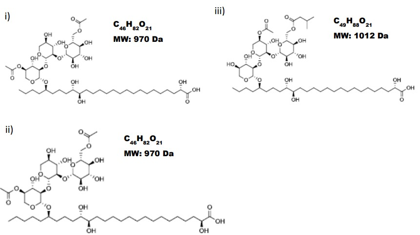
AM-1 is a mixture of structurally related long-chain glycolipid congeners, with the glycolipids contributing ≥ 93% on dry weight basis, calculated as sodium salts. The three main components are provided below in Figure 1.
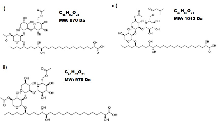
The remaining components are congeners of the major components, sharing the saturated C26:0 fatty acid and the glucopyranosyl-(→)-xylopyranosyl-(→)- xylopyranosyl trisaccharide moiety but differing in the acylation pattern, i.e. the number and position of acyl groups attached to the sugar units; the acyl groups are usually acetate and isovalerate.
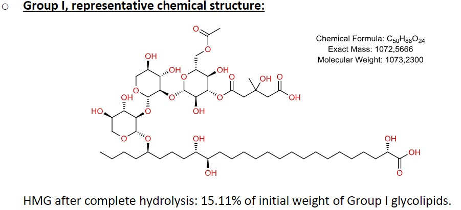
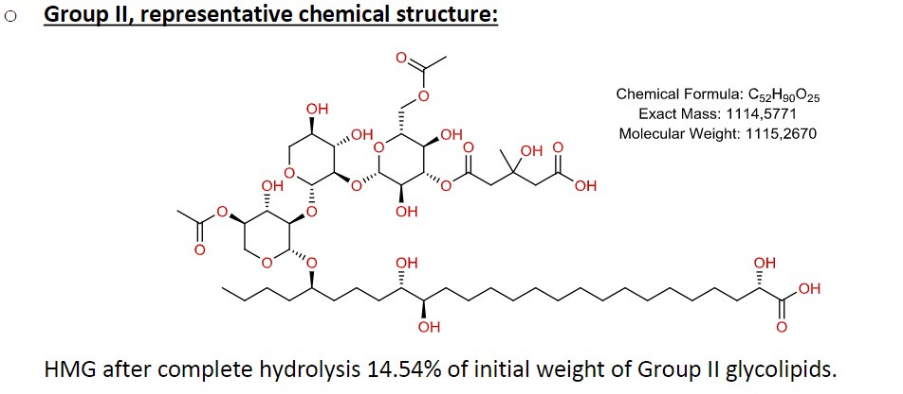
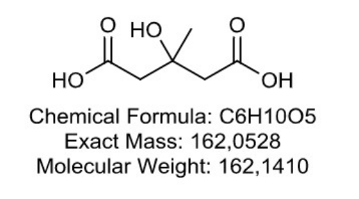
For certain batches of AM-1 the minor component 3-hydroxy-3-methylglutarate (HMG) has been noted as an acyl unit instead of acetate and isovalerate; the average calculated concentration of HMG is < 1.5% (w/w).
AM-1 has the following normalised glycolipid profile (Figure 2) as determined by high-performance liquid chromatography – mass spectrometry (HPLC-MS):

The AEJEG was satisfied that the analytical results for five non-consecutive batches of commercially representative AM-1 demonstrated conformance to specifications and consistency of AM-1 manufacturing.
An updated specification was provided to the AEJEG after the original Application submission to align with the EFSA approved Glycolipids (E246) Specification.
2.3. Specification
The Applicant highlighted that the European Commission had not found it necessary to include the glycolipid profile in the specifications. Furthermore, the Applicant raised that the OECD Technical Guideline 105 on Water Solubility could not be followed as AM-1 is comprised of a mixture of glycolipids. As a consequence, AM-1 is also a biosurfactant which does not exhibit classical molecular solubility and this usually makes it impossible to determine maximum solubility of the compound. The solubility was instead verified at a fixed concentration (10 g/L) in what the Applicant refers to as a ‘shake-flask’ test to visually confirm solubility. The solution properties of AM-1 were then corroborated by turbidity measurements using the same solution, which the Applicant noted is an approach accepted by EFSA (EFSA, 2021). The Applicant defined the solubility close to application of the product and to ensure that the value reported is of practical relevance for food manufacturers. This value is complemented by the determination of turbidity of the same aqueous solution, guaranteeing no cloudy solutions are accepted and avoids subjective appraisal of turbidity, which would lead to non-reproduceable results.
The AEJEG accepted the method used by the Applicant, and it was noted overall the AEJEG considered the product to be water miscible.
The final product was tested for Coliforms instead of Enterobacteriaceae to verify good hygienic practice in the specification of AM-1 (glycolipids). With the applicant stating that “both methods are common parameters to account for good hygienic practice and are, in practice, regarded to be equivalent”. The Applicant further stated “Coliforms belong to the family of Enterobacteriaceae and are proposed to be monitored with an even stricter value of ≤ 3 MPN/g. We are convinced that the Most Probable Number (MPN) method is suitable because it is performed in a liquid solution rather than a dry powder. The liquid broth represents the intended use of AM-1”.
The free fat content listed as <2% was clarified to refer to free fat that did not include the fatty acids which are part of (and bound in) the glycolipids; fatty acids which are part of (and bound in) the glycolipids are included in the specified minimum content of 93% for glycolipids. The AEJEG suggested that the specification could instead be labelled as ‘free fat content’ to avoid confusion. The AEJEG also suggested that this issue should be flagged to risk managers for their consideration.
2.4. Information on particle size, particle size distribution and other physicochemical characteristics
AM-1 is not an ‘engineered nanomaterial’ as defined in Article 3 2. (f) of assimilated Regulation (EU) 2015/2283 of the European Parliament and of the Council on novel foods. Laser diffraction analysis (LDA) provided the particle size distribution, and scanning electron microscopy (SEM) was used to verify the laser diffraction results and, additionally, investigate if nano-scale material is present as clusters or adsorbed onto larger-scale particles. Both techniques were carried out in parallel on six representative batches produced in 2019 or 2020.
LDA shows that the mean particle size was 42.8 µm. The values Dx (10) (the size below which10% of the material is present) showed clearly that 10% of the samples had a size of 10.3 µm or less (the average Dx (10) of all samples is 14.9 µm). SEM confirmed that AM-1 consists essentially of spherical particles averaging a few tens of micrometres in size, which corresponded with the mean particle size determined by LDA. Only a few small particles could be seen on the large crystals, which have particle sizes of 200 nm or more.
2.5. Manufacturing Process
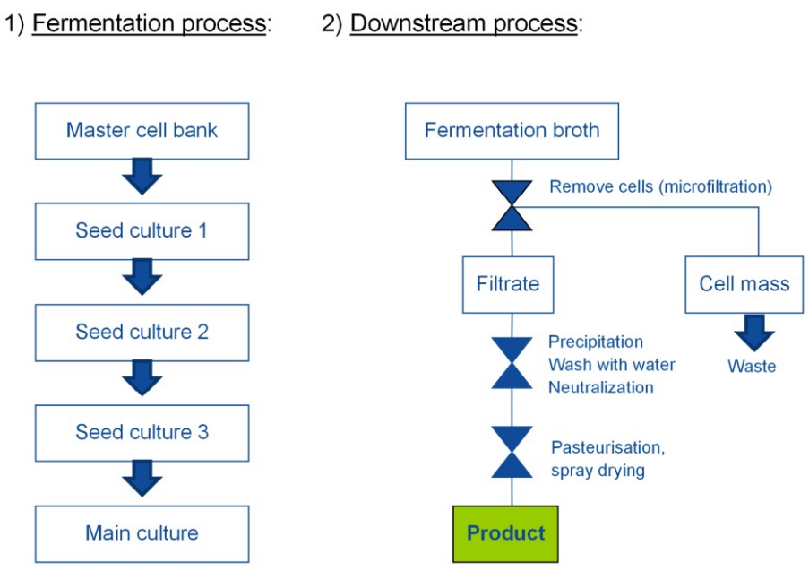
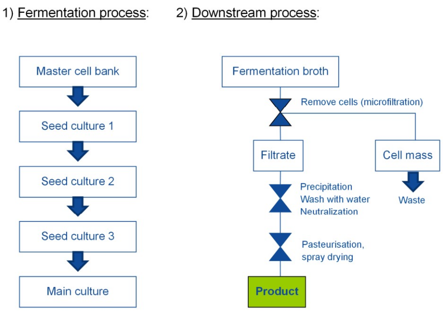
Fermentation of Dacryopinax spathularia strain MUCL 53181 is conducted using glucose as the carbon source in an aerobic submerged culture. Starting from a cryogenic working cell bank (WCB), a series of seed cultures are grown in shake flasks and then in bioreactors with increasing volumes to obtain sufficient living fungal cells to inoculate the main culture. This main culture is conducted in fed-batch mode, with start and feed media both consisting of glucose and small amounts of yeast extract (commercial extract of autolysed Baker’s yeast, Saccharomyces cerevisiae). The culture is maintained at 30°C for several days until maximum titre of glycolipids is achieved. The feed is stopped, and the cells are further cultivated until no free glucose is found in the culture medium.
Fungal cells are quantitatively removed by microfiltration, followed by acidic precipitation of the glycolipids. The precipitate is washed with water and neutralized using sodium hydroxide solution. Spray or freeze-drying leads to the final product as an off-white, water miscible powder. The presence of any remaining intact fungal cells from the source organism Dacryopinax spathularia is excluded. This is assured by the design of the microfiltration step and has been confirmed by viable fungal cell count (using preferred growth conditions for Dacryopinax spathularia strain MUCL 53181) as well as microscopic control of representative batches.
The Applicant provided an overview of the manufacturing process as provided in Figure 3. The production process follows Good Manufacturing Practices (GMP) and Quality Management compliant with Food Safety Scheme FSSC22000, including a Hazard Analysis and Critical Control Point (HACCP).
The Applicant later provided further information stating “microscopic investigations did not reveal any cell[s] (viable or not) and confirmed the absence of any viable fungal cell[s] in the product, therefore strongly suggesting that no cells of the production organism are present in the commercial product AM-1. These study results corroborate the conclusion based on the production process, which physically excludes the presence of cells by a series of filtration steps, including microfiltration.”
Full reports and figures were provided by the Applicant to support these claims and the AEJEG deemed these acceptable.
After pasteurisation, the product is spray dried. The resulting powder is then directly mixed in a powder mixer and packaged in a clean room. Cleaning procedures and environmental monitoring are in place to reasonably prevent any microbial re-contamination.
The Applicant claimed the risk of contamination with pathogenic microbes after pasteurisation is rated to be negligible. Nevertheless, appropriate controls are included in the specification of the material to verify microbial purity for each batch.
2.6. Presence of impurities
The Applicant stated that no impurities other than those listed in the specification (protein, fat, water, and sodium chloride) have been observed or are expected. The Applicant provided analytical results regarding the presence of heavy metals and microorganisms for five non-consecutive batches.
The Applicant stated by-products are not expected based on the manufacturing process as the AM-1 powder enters the powder mixer directly from the spray-dryer before this it is pasteurised and concentrated. The efficiency of this pasteurisation step was validated by total plate count of a minimum n=5 batches. Sampling is done at the beginning, three intermediate time points and end of the packaging process, and sub-samples are combined into one composite sample.
The composite sample is divided in two parts, and each is analysed for a full set of parameters as listed on the Specification in two independent laboratories; two-class attribute plans are used for microbial detection and enumeration.
The AEJEG had concerns that as the product is always dispersed in water for application in the form of a stock solution, there may contamination with pathogenic bacteria such as Listeria. The Applicant responded that due to AM-1 being applied as a 1% aqueous stock solution, microorganisms susceptible to AM-1 would be killed immediately during stock solution preparation. Therefore, there is no hazard identified regarding Gram-positive food pathogens like Listeria, Bacillus or Staphylococcus.
AM-1 has reduced or no antimicrobial efficacy against Gram-negative pathogenic bacteria. The absence of E. coli, other coliform bacteria and Salmonella is confirmed by microbiological sampling and testing. To include a generic (“horizontal”) test for aerobic bacteria, which might potentially not grow in the specific (“vertical”) assays, total aerobic plate count (TAMC) is performed. Further, AM-1 is efficacious against vegetative fungal cells, but it is not sporicidal. To address the presence of fungal spores, total yeast / mould count (TYMC) is included.
The Applicant therefore concluded that their setup of production control including pasteurization and a cleanroom packaging, besides additional analytical data, ensures the high microbial purity of AM-1.
Heavy metals arsenic (As), lead (Pb), nickel (Ni), cadmium (Cd) and mercury (Hg), thresholds have been provided within the specification. Furthermore, analytical results for each batch have been given in the Certificate of Analysis (COA), the levels permitted in the specification are given in Table 2.
Based on the composition and the method of manufacture, the Applicant stated the presence of the following impurities can be excluded; alcohol (ethanol), benzene, 1,4-dioxan, dioxins, ethylene oxide, formaldehyde, Genetically Modified Organisms (GMOs), gluten, glycol ethers, nitrite, nitrosamines and pesticides.
The lyophilised fungal fermentation broth (two independent batches) and the glycolipid product AM-1 (two independent batches) were both analysed for the presence of mycotoxins, and no mycotoxins were detected. Methods of analysis included HPLC, HPLC with tandem MS (HPLC/MS-MS), HPLC with a fluorescence detector (HPLC-FLD) and liquid chromatography with a triple quadrupole MS (LC-MS/MS).
Details of the 4 batches that were analysed for potential mycotoxin content at the two specialised analytical laboratories, the test reports, limits of quantitation and certificates of analysis were provided and reviewed by the AEJEG, who concluded these were acceptable.
The Applicant claimed, based on a literature research and results of their in-house analytics, the presence of antibiotics has been excluded. Small molecular weight components other than glycolipids and carotenoids, which are the only other known secondary metabolites that are produced by strain MUCL 53181, were searched for extensively using HPLC-MS, HPLC with ultra-violet spectroscopy (HPLC-UV) and HPLC with an evaporative light scattering detector (HPLC-ELSD), however no traces were found. Furthermore, the anti-bacterial bioassays showed only moderate anti-bacterial activity in the presence of the glycolipids. Removal of the glycolipids from the fermentation broth (e.g. by acidic precipitation) also removed the anti-bacterial activity. Therefore, all credible evidence available supports the absence of any medically or clinically relevant antibiotics in the fermentation broth of strain MUCL 53181.
Two fermentation samples of Dacryopinax spathularia strain FU50088, four AM-1 batches produced at pilot-scale and four commercial batches produced in industrial-scale were subjected to analysis by the dereplication method at a concentration of 1 g/L or 10 g/L respectively. Four representative fungal antibiotics had been previously used to determine the detection limit.
The mass spectra of the antibiotics and all chromatographs of all samples showing all detector signals were provided to the AEJEG.
The signals visible in the different chromatogram traces are summarised in Table 3. In chromatograms of the positive electrospray ionization (ESI) trace some signals are system-intrinsic signals from the LCMS (artefacts). These were detectable in all analyses recorded with this LCMS system and method, including blank runs. Therefore, the Applicant concluded these peaks did not represent components in the analysis samples.
The assigned signals listed are not present in all the samples. However, if there were signals at a given retention time and detector, the identified metabolites were identical across all samples.
Some residual lipids were detectable in the final product batches, alongside minor amounts of polar metabolites in the fermentation samples. The detection limit for fungal antibiotics for the applied methodology was found to be 1 mg/L or below, and a MS detector was used as reference for exclusion of the presence of antibiotics or other low-molecular weight components.
Considering the limit of detection (LOD) of <1 mg/L and the sample concentrations of max. 10 g/L, compounds present at 0.01% of dry weight or more are expected to be found.
During project development, various AM-1 glycolipids had been isolated as pure components by preparative chromatography. No metabolites other than glycolipids were ever observed.
The AEJEG determined this information was acceptable.
2.7. Stability, reaction and fate in foods to which the additive is added.
2.7.1. Dry powder
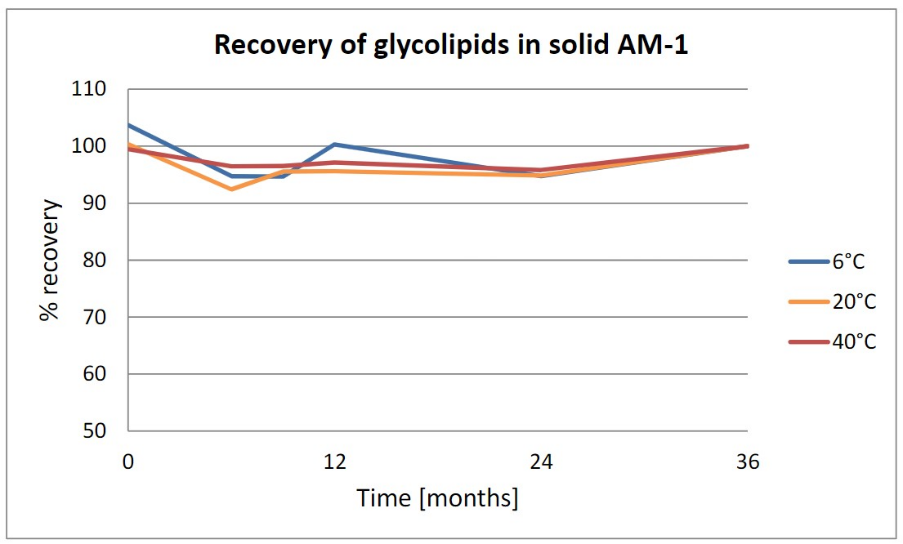
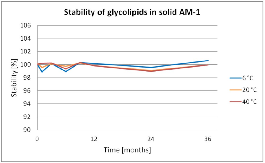
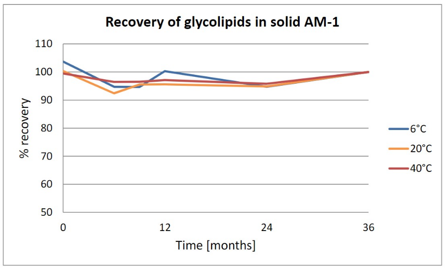
AM-1 as a dry powder stored in a closed container is stable at temperatures of 40°C and at an ambient temperature of 20 °C for at least three years without any detectable degradation or modification of its own composition as shown in Table 4 and Table 5.
Full details of this study and the analytical sample preparation, dilution schemes, LCMS setup and determination of stability in [%] from the glycolipid profile were submitted to the AEJEG via an RFI response. The AEJEG determined that the study design and the results were acceptable.
Analytical Results
Physical Stability
Colour and appearance of the samples were assessed over 36 months; no visual changes of the test item were observed at any storage temperature and time point.
Recovery (glycolipid quantification)
Percent recovery determined by LCMS-based glycolipid quantification did not show any change or loss in the glycolipid content at any temperature during the test period. Figure 4, raw data was provided by the applicant to the AEJEG.
Chemical stability (Glycolipid profile)
The AM-1 glycolipid profile was found unchanged at all temperatures throughout the test period of 36 months. Figure 5, raw data was provided to the AEJEG by the applicant.
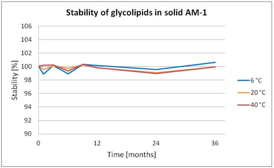
No degradation or hydrolysis of glycolipids, and no change in physicochemical properties was found. The Applicant therefore concluded that AM-1 as a dry powder was stable at 6 °C, 20 °C, and 40 °C for at least 36 months.
When stored as a dry powder in a closed container (HDPE drums) at temperatures of 40°C and below, no detectable degradation or modification of its composition occurs. This was overviewed in the recommended storage conditions as outlined in the Safety Data Sheet, which was provided to the AEJEG by the applicant.
The applicant stated that “Parameters such as light, or oxygen have no or negligible effects on the glycolipids since the fatty acid and the trisaccharide do not offer a target for a chemical reaction with oxygen or light such as autoxidation of the fatty acid. As no UV chromophore or light-sensitive chemical structure is present, no impact of light or oxygen on storage stability is expected.”
The AEJEG determined this was acceptable.
2.7.2. Aqueous solution
Long term stability tests over 3 years were carried out on the product in aqueous solution. Details of the study set up were provided to the AEJEG and determined to be acceptable.
Results
Physical Stability
Colour and viscosity of the samples were assessed over 36 months, and the findings are summarised in; Table 6, Table 7, Table 8, Table 9 and Table 10.
The AEJEG reviewed the information in Table 3 (Stability) in addition to the detailed explanation of the data and photographs of the colour changes (Table 4) provided by the Applicant and determined this was acceptable.
Chemical Stability
AM-1 is a mixture of glycolipids, all belonging to the same structural class and differing in the number and type of small organic acids attached as esters to the sugar units. Over time partial hydrolysis of these esters (deacylation) occurs.
Graphs and raw data were provided to the AEJEG, who agreed with the Applicant that degradation of the glycolipid core structure (i.e. deglycosylation) had not been observed in any of the test samples.
The Applicant stated that “Non-acylated and monoacylated glycolipids (herein referred to as “deacylated glycolipids”) also exhibit antimicrobial properties, although with slightly lower potency. Therefore, a stability value in this assay of e.g. 90% does not correspond to a loss of 10% of antimicrobial activity. Instead, reduction of antimicrobial activity is significantly lower than % hydrolysis”.
Conclusions
At 6 °C, no change of the AM-1 glycolipid profile was detectable for the first twelve months. After 24 and 36 months, only very minor hydrolysis of acyl units (<5%) was found in all samples.
Physical effects such as sedimentation were found to be fully reversible by stirring at 60°C for 10-15 min so were not regarded as a sign of instability. Hydrolysis of AM-1 solutions in pure water was found to occur in a time-dependent manner above 6°C.
In conclusion, AM-1 solutions in pure water were regarded as chemically stable for 1-2 months when stored at room temperature. After 3 months or longer storage periods, deacylation of more than 5% of the glycolipids had occurred, leading to a modified but still biologically active product. Degradation of the glycolipid core structure had not been observed. Increased viscosity and sedimentation were observed but were regarded as reversible effects.
At 40 °C, hydrolysis of ester units proceeds faster than at ambient temperature. After 1 month, 5-10% of the glycolipids had been hydrolysed to mono- and deacylated glycolipids. The hydrolysis rate was similar for all AM-1 concentrations, being circa. 55-70% within 12 months. With increasing incubation time, the rate was reduced to 15-20% for the 12-25 months period. After 25 months a plateau was reached, and further hydrolysis could be detected only to a minor extent.
In conclusion, at 40°C AM-1 solutions in pure water were regarded as chemically stable for 2-7 days when stored. At the end of the test period, non-esterified glycolipids are the predominant species, ensuring at least a basic antimicrobial effect. Other degradation products were not observed in significant amounts. Visually, darkening of the samples was observed for all concentrations. Sedimentation occurred at 1.5% and 10% AM-1 (12 months), solidification at 10% AM-1 level only (24 months).
The Applicant recommended that AM-1 solutions be stored under refrigerated (4 – 8°C) conditions for up to 1 year, e.g. for use in a laboratory environment. Longer storage periods of AM-1 solutions in industrial beverage production would not be required, are not feasible, not intended and not foreseen. Instead, AM-1 powder would be dissolved into a stock solution on the day of production.
2.7.3. Beverage Applications
The stability of AM-1 in beverages was tested in 110 commercially obtained beverages chosen from different categories, including carbonated soft drinks (CSD), fruit drinks (FD), enhanced waters, sport drinks, energy drinks, syrups and various ready-to-drink teas and stored for 3 months at ambient temperature. “Analysis was conducted via HPLC-MS due to technique being able to differentiate between individual glycolipid congeners of the glycolipid mixture”. Degradation of the glycolipids to its principal components: glucose, xylose and long chain fatty acids (LCFA) did not occur in any beverage. Details of the study design and analysis were provided to the AEJEG and determined to be acceptable.
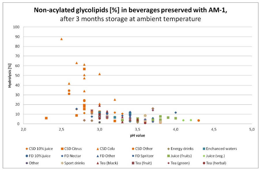
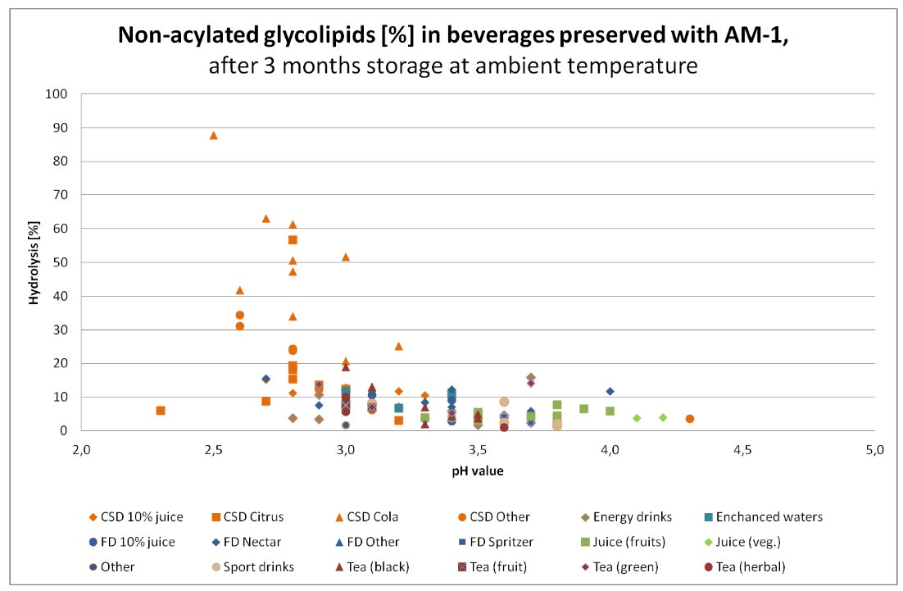
AM-1 was determined to be stable in beverage applications of various pHs under typical storage conditions, with the hydrolysis of the ester moieties of the glycolipids dependent on the pH value and cloudiness of the individual beverage. Beverages with low pH generally showed a higher degree of ester hydrolysis compared to cloudy fruit juices, and beverages with a pH greater than 3.5 showed low degrees of ester hydrolysis. Figure 6 in which a percentage of 100% corresponds to a complete deacylation of the glycolipid mixture, raw data was also provided to the AEJEG for all substances.
Results
The ratio of deacylated glycolipids in a glycolipid mixture is a measure for AM-1 stability in beverages: the lower the ratio of non-acylated glycolipids the more stable the glycolipid mixture. The Applicant claimed the preserving properties of partially deacylated glycolipids are still very good but slightly reduced compared to the initial material. This was shown by antimicrobial tests; however, the data were not reported in the dossier submitted to the FSA.
Hydrolysis of ester moieties of the glycolipids was found, to a degree, to be dependent on the pH value and cloudiness of the beverage: those with a pH >3.5 showed only low degrees of ester hydrolysis compared to carbonated soft drinks of lower pH.
The Applicant highlighted how it was important to note that “even after deacylation, the glycolipid structures as such remain intact. There have been no findings of deglycosylation of glycolipids in the analysed beverages, i.e. there is no decomposition of glycolipids into long-chain fatty acids (LCFA) and sugar units”.
The Applicant also confirmed that the long-chain glycolipids of AM-1 were not prone to oxidation due to the fatty acid backbone being fully saturated, and no other chemical units prone to oxidation being present. The applicant stated that the presence of oxygen will impact microbial growth and risk for a beverage, but not the stability of AM-1.
Furthermore, the long-chain glycolipids were not degraded by the influence of light and do not contain UV/VIS chromophores such as unsaturated fatty acids or aromatic rings and were therefore not expected to be degraded by the impact of sunlight.
2.7.4. Hydrolysis in aqueous solutions
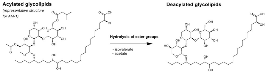
The Applicant claimed the hydrolysis of ester moieties proceeds in a time, pH, and temperature dependent manner, shifting towards deacylated glycolipids as shown in Figure 7. The hydrolysis products are acetate and isovalerate which are normal constituents of the human diet and if absorbed are rapidly eliminated and may be metabolised to CO2.
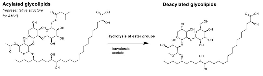
The Applicant stated that longer incubation times, lower pH values (<3.5) and higher temperatures (≥40°C) all favour the hydrolysis of acetyl and isovaleryl ester moieties of the AM-1 glycolipids, increasing the ratio of deacylated glycolipids present in the mixture. The best antimicrobial efficacy is observed for the acylated glycolipids, and partial or full deacylation reduces the potency. However, even the fully deacylated long-chain glycolipids maintain very similar antimicrobial properties, with only slightly (2-fold) higher Minimum Inhibitory Concentrations (MICs).
At the point in time when partial deacylation has occurred in beverage applications, it is expected that the residual viable cell count is already zero or close to zero (e.g. only very few remaining spores). Therefore, the conclusion is that the antimicrobial properties of the deacylated glycolipids are suitable for their intended use.
There are no degradation products of safety concern associated with AM-1 for both the bulk material or when formulated in low pH beverage application under the typical storage and use conditions. The hydrolysis mechanism and resulting products are expected to be the first step of degradation in the gastrointestinal tract following ingestion of AM-1 and is consistent with experimental results in simulated gastric / intestinal fluid and in rodents.
The hydrolysis of AM-1 containing the 3-hydroxy-3-methylglutarate (HMG) acyl unit has been covered in the safety assessment of exposure to this constituent and is presented within the toxicity section.
Chemically, AM-1 glycolipids can be described as biosurfactants having a hydrophobic long-chain fatty acid backbone and hydrophilic trisaccharide unit. Therefore, physical interactions with the beverage matrix such as adsorption effects are expected to occur and explain the different dosages required for AM-1 to be applied in different beverage matrices. However, no chemical reactivity has been observed for the glycolipids and no chemical interaction is expected based on the low reactivity (almost inertness) of the saturated chemical structures of the components. This is supported by the stability data for AM-1 in beverages and in solution, where a change of the glycolipid composition has been shown due to partial deacylation, but the full amount/concentration of the product was shown to be persistently present throughout the product’s shelf life.
2.8. Existing authorisations and previous risk assessments
AM-1 has been authorised for use in Europe, Canada, Australia and New Zealand. It has been assessed as GRAS (‘generally recognised as safe’) within the United States. The Applicant confirmed that the same studies had been submitted to EFSA, Health Canada (HC) and Food Standards Australia New Zealand (FSANZ), with differing Acceptable Daily Intakes (ADIs) established by these regulating bodies.
2.8.1. Australia and New Zealand (FSANZ)
AM-1 has been authorised in Australia and New Zealand by the Food Standards Australia New Zealand (FSANZ) in 2021 in Amendment No. 198 to the Australia New Zealand Food Standards Code.
FSANZ had assessed an Application from LANXESS Deutschland GmbH to amend the Australia New Zealand Food Standards Code (the Code) to permit the use of a long-chain glycolipid mixture [AM-1] derived from Dacryopinax spathularia (‘jelly mushroom glycolipids’) for use as a food additive (preservative) in non-alcoholic beverages.
The maximum permitted levels of jelly mushroom glycolipids to be permitted in the associated non-alcoholic beverages categories are:
-
100 mg/kg for fruit and vegetable juices and fruit and vegetable juice products
-
50 mg/kg for water based flavoured drinks;
-
20 mg/kg for Formulated Beverages;
-
10 mg/kg for coffee, coffee substitutes, tea, herbal infusions and similar products;
-
100 mg/kg for non-alcoholic beer.
In a 90-day Good Laboratory Practice (GLP)-compliant study in Sprague Dawley rats, no adverse effects were found at the highest dose administered, equivalent to 1201 and 1423 mg/kg bw per day in male and female rats, respectively. In a 90-day GLP-compliant study in Beagle dogs, a significant reduction (relative to that of controls), in group mean cumulative bodyweight gain was seen in female dogs, with a corresponding but non-significant reduction in group mean food consumption, at 1000 mg/kg bw per day. This observation was considered to be adverse by FSANZ. FSANZ considered that the No Observed Adverse Effect Level (NOAEL) for this study was 500 mg/kg bw per day.
The ADI was based on the lowest NOAEL identified in animals, 500 mg/kg bw per day in Beagle dogs. FSANZ applied an uncertainty factor (UF) of 10 for extrapolation from animals to humans, an UF of 10 for variability between humans, and an UF of 3 allowing for extrapolation from a subchronic study to chronic exposure, for a total UF of 300. An uncertainty factor of 3, rather than 10, was selected because there was a clear NOAEL at 500 mg/kg bw per day, and the effect is minimal at twice that value, although the dogs were at an age when growth is rapid and energy requirement is high. The ADI was established by division of the lowest NOAEL (500 mg/kg bw per day) by the total UF (300), approximately equalling 1.6, and rounded to 2.0 mg/kg bw per day.
FSANZ approved the use of ‘sweet osmanthus ear glycolipids’ which was considered a more appropriate term for AM-1 as a food additive in non-alcoholic beverage categories at the maximum permitted levels sought. The Amendment No. 198 to Food Standards Code permitting the use of AM-1 was gazetted in Australia (No. FSC 139, 26 March 2021) and published in the New Zealand Gazette.
2.8.2. Canada (HC)
Health Canada’s Food Directorate received a food additive submission seeking approval for the use of long-chain glycolipids [AM-1] from Dacryopinax spathularia MUCL 53181 as a preservative in certain standardized and unstandardised non-alcoholic beverages. This request was approved in June 2022. The requested maximum use levels range from 25 parts per million (ppm) to 100 ppm in the finished beverage.
The Food Directorate concluded that information related to the safety and efficacy of long-chain glycolipids from D. spathularia MUCL 53181 supports its use as an antibacterial agent and an antifungal/antimycotic agent in the beverages of interest in the below Food Categories.
Health Canada established an ADI of 2.5 mg/kg bw, noting its use as a preservative as proposed does not pose any chemical, microbiological, nutritional or toxicological safety concerns.
The ADI established by Health Canada is defined as a “Regulatory ADI”, a term which Health Canada have used themselves. Based on the existing data, Health Canada had established an ADI that was sufficiently high to support the requested Application of glycolipids for the following food categories:
The results of the premarket assessment support the safety and efficacy of long-chain glycolipids from D. spathularia MUCL 53181 for use as a preservative as set out in the table above (Table 11). Health Canada therefore proposed to enable the uses of this new food additive as shown above. The Food Inspection Agency Canada (FIAC) also approved the synonym Glycolipids for labelling in Canada.
The Applicant highlighted to the AEJEG that “the MPLs [Maximum permitted level] approved by Health Canada are significantly higher than those requested for in the UK and include fruit juices as a category which has not been applied for in the UK. Health Canada considered a Regulatory ADI of 2.5 mg/kg bw to be sufficiently high to approve the MPLs as listed in the above table” (Table 11).
The Applicant also reported to the FSA that Health Canada has confirmed that they would revise the ADI if they received Applications for higher MPLs and/or additional food categories. Health Canada also confirmed that they would align the ADI of glycolipids with an ADI established by JECFA as soon as it is available.
The AEJEG was not provided with confirmation in writing of the actual statements made by Health Canada. Following discussion, the AEJEG noted a difference in interpretation regarding the information provided by Health Canada. The AEJEG disagreed with the Applicant that the Health Canada ADI was linked to the proposed use levels and, subsequently, that they are subject to change.
2.8.3. EFSA
In 2021 the EFSA Panel on Food Additives and Flavourings (FAF) provided a scientific opinion on the safety of long-chain glycolipids from Dacryopinax spathularia (also called AM-1) as a food additive.
Considering the available dataset the Panel established an ADI of 10 mg/kg bw per day based on a range of NOAELs between 1,000 and 1,423 mg/kg bw per day (the highest doses tested), from the reproductive and a prenatal developmental toxicity study in rats and 90-day studies in rats and dogs (Bitzer et al., 2018).
At the proposed maximum use levels, the exposure estimates ranged at the mean from 0.01 to 1.07 mg/kg bw per day and at the p95 from 0 to 3.1 mg/kg mg/kg bw per day. This data set was reported by EFSA (2021) and although it is noted that the range for the p95 starts at a lower value than that for the mean this was all the data provided by the Applicant.
At the proposed typical use levels, the exposure estimates ranged at the mean from < 0.01 mg/kg bw per day to 0.23 mg/kg bw per day and at the p95 from 0 to 0.64 mg/kg bw per day.
The EFSA Panel noted that the highest estimate of exposure of 3.1 mg/kg bw per day in toddlers is within the established ADI of 10 mg/kg bw per day and concluded that the exposure to long-chain glycolipids from Dacryopinax spathularia did not raise a safety concern at the uses and use levels proposed by the Applicant.
Commission Regulation (EU) 2022/1037 of 29 June 2022 amending Annex II to Regulation (EC) No 1333/2008 of the European Parliament and of the Council and the Annex to Commission Regulation (EU) No 231/2012 as regards the use of glycolipids as a preservative in beverages came into force on the 20 July 2022.
2.8.4. United States
AM-1 had been assessed as generally recognised as safe (GRAS) in accordance with section 201(s) (21 U.S.C. § 321(s)) of the Federal Food, Drug, and Cosmetic Act (21 U.S.C. § 301 et. Seq.) of the United States of America. The Applicant provided further details of this evaluation. However, the AEJEG considered as the system works differently in the USA, and as it possible for companies to self-certify an ingredient as GRAS as opposed to this being determined by the FDA, it was agreed that the GRAS status would have limited impact on the AEJEG conclusions.
2.8.5. Applicant’s comments on differences of the evaluation between FSANZ and EFSA.
The Applicant provided a comment to the AEJEG regarding the differences between the FSANZ and EFSA evaluation conclusions, “FSANZ applied a NOAEL of 500 mg/kg bw per day (instead of 1,000) and a safety factor of 300 (instead of 100) because of their interpretation of the toxicological data. EFSA did not concur with FSANZ’s assessment and determined the NOAEL to be 1,000 mg/kg bw per day and saw no need to deviate from the usual safety factor of 100”.
2.8.6. Joint FAO/WHO Expert Committee on Food Additives (JECFA)
the FSA/FSS note that at the 100th meeting in June 2025, JECFA considered and assessed the safety of glycolipids. The Joint FAO/WHO Committee concluded that the estimated dietary exposures did not raise any safety concerns.
2.9. Proposed normal and maximum use levels.
The Applicant proposed AM-1 be used in the following categories of food (Table 12) according to Annex II Part D of Regulation (EC) No 1333/2008 on food additives with the following use levels:
2.10. Dietary exposure assessment and data on dietary sources
The Applicant submitted dietary exposure estimates for glycolipids (AM-1) based on the proposed maximum and normal use levels using EFSA’s Food Additive Intake Model (FAIM). Calculations were performed on 11th January 2022, with an ADI of 0-10 mg/kg bw per day based on the 1000 mg/kg bw per day NOAEL from the 90-day oral toxicity study in Beagle dogs (Bitzer, Henkel, Nikiforov, Rihner, & Henderson, 2017) and a safety factor of 100 for AM-1 used to calculate the margin of safety. This original exposure assessment is provided in Appendix 1.
It was noted that when running these calculations, the values calculated by the AEJEG were higher than those presented by the Applicant. The AEJEG considered this could be due to the version of the model used by the Applicant in their submission or the way they have assigned the use levels to the specific food commodities.
The AEJEG agreed the recalculated exposures (performed by the AEJEG) should be utilised for the Committee Advice Document on AM-1.
The recalculated dietary exposures have been presented below based on the following use levels and inputs into the EFSA’s FAIM tool revision. 21 (calculations performed in June 2024):
Based on the maximum use levels, dietary exposures were provided in Table 13.
Based on the typical use levels, dietary exposures were provided in Table 14.
These intakes were marginally higher than the estimates provided by the Applicant and the AEJEG considered this as the worst-case scenario. It was also noted that intakes for UK consumers (based on the Applicant’s submission) were lower than the intakes presented above for the critical EU diet for each corresponding consumer group. The AEJEG noted that the reliance on non-UK exposure estimates added a degree of uncertainty that was assumed to lead to an overestimate of exposure.
The Applicant also indicated that intakes may be overestimated due to it not being possible to distinguish between alcohol-containing and alcohol-free variants within category 14.2.1 (beer and malt beverages). However, the AEJEG also considered that the consumption patterns of non-alcoholic and alcoholic beverages should be viewed as similar. Nevertheless, it should be noted that this food category does not contribute to the exposure of the critical consumer (toddler).
The highest exposure based on the maximum use level was 3.08 mg/kg bw per day (max 95th Percentile for toddlers). However, this intake is reduced to 0.64 mg/kg bw per day when considering typical use levels, and the AEJEG agreed this reflected a more realistic scenario when considering long-term/lifetime exposures.
Exposure was compared to the ADI agreed by the AEJEG of 3.3 mg/kg bw per day, based on the NOAEL of 1000 mg/kg bw per day for the 90-day rat study (LPT, 2015) with an uncertainty factor of 100 x 3 (to account for the subchronic nature of the study).
The AEJEG noted intakes for all consumers are below this level for both the typical and maximum use levels. It is recognised that the maximum use level scenario is highly conservative, and typical use is more representative of lifetime exposure.
2.10.1. Exposure to contaminants
For inclusion in the Committee Advice Document, the AEJEG performed a dietary exposure calculation for the heavy metals present in the product.
Maximum exposures to toxic elements (arsenic, lead, nickel, cadmium and mercury) from the use of these glycolipids were estimated from their maximum limits provided in the specification and the maximum 95th percentile exposure for the maximum use levels for toddlers. Calculations and reference points for each element have been provided in Table 15.
The AEJEG noted that the reference points for arsenic were based on carcinogenicity and that EFSA had indicated the MOE should be 10,000, and for lead the MOS/MOE should be 1 or greater with the reference point based on a BMDL01 of 0.5 μg/kg bw per day (EFSA CONTAM Panel, 2010).
Arsenic levels were considered by the AEJEG as a potential concern as the margin of exposure (MOE) for arsenic was 2,500, where a normal value of 10,000 would be expected when based on the Benchmark Dose Level (BMDL). However, Members acknowledged that other agencies did not consider this to be a significant issue. Levels of the other metals did not give rise to any safety concerns.
2.10.2. Exposure in other sources
Glycolipids such as AM-1 are part of the normal human diet, the most abundant sources being eggs, dairy and cereal. Sphingolipids are important components of the cell membrane, glycerolipids and phosphoglycerolipids (both having shorter lipid chain lengths than AM-1) are abundant membrane lipids and as such part of the human diet.
The Applicant was asked to provide clarification whether the use of the additive will produce exposure to any other toxicologically relevant substances, e.g. from potential residues of degradation products, reaction products, and contaminants. The Applicant replied that “the potential degradation products of AM-1 are simple components such as small organic acids, glucose and fatty acids, all of which are of no toxicological concern”.
2.11. Biological and toxicological data
2.11.1. Historical Consumption
Glycolipids are part of the normal human diet, with the most abundant sources identified as eggs and dairy products, cereals and soybeans (Leray, 2015). The fruiting bodies of the producer organism (Dacryopinax spathularia) are edible and have traditional use as food in many countries in Asia and Africa. Furthermore, a field guide to foraging mushrooms in Oregon (USA) includes a recipe with Dacryopinax spathularia alongside identification of this edible mushroom (Meuninck, 2017), showing use as food in the USA.
Dacryopinax spathularia is listed in the Food and Agriculture Organization of the United Nations (FAO) compendium on edible mushrooms (Boa, 2004) with proven food use documented in China (Zhishu et al., 1993), Indonesia (Ellen, 2008) and Cameroon (van Dijk et al., 2003).
The Applicant claimed the presence of AM-1 as a constituent in collected fruiting bodies of Dacryopinax spathularia had been confirmed by HPLC-MS analyses. Further, AM-1 was also found in small fruiting bodies of the producer strain MUCL 53181 grown on wood substrate in the laboratory.
2.11.2. Applicant’s Safety Assessment of AM-1 Production Organism
The safety of the AM-1 production organism Dacryopinax spathularia MUCL 53181 was assessed by the Applicant utilising scientific procedures outlined by Pariza and Johnson (2001) for safety evaluation of microbial enzyme preparations used in food processing. Using the paradigm, the AM-1 ingredient may be considered the “test article” that is produced by a mushroom fermentation culture, which is comparable to an enzyme preparation produced by a microbial culture.
Based on the Pariza and Johnson (2001) Decision Tree Analysis of AM-1 Production Organism the Applicant concluded that Dacryopinax spathularia MUCL 53181 is considered safe for use in the production of AM-1 as an ingredient for human consumption.
2.11.3. Toxicological studies (Toxicokinetics and Toxicity)
Literature papers, as well as study reports were provided; all safety studies have been performed by LANXESS or its subsidiaries using glycolipids (AM-1) as a test material.
2.11.4. Absorption, Distribution, Metabolism and Excretion (ADME) Profile of AM-1
Male and female Sprague Dawley rats aged 8-10 weeks were used to evaluate the pharmacokinetics, excretion balance (mass balance as a measurement of test-article equivalents in excreta samples), and tissue distribution of [14C]-AM-1 and [14C]-Long Chain Fatty Acid (LCFA) equivalents following single or repeated administration. The study was performed in compliance with GLP Regulations, and the study protocol was designed in general accordance with U.S. FDA Redbook Guideline Redbook II Guidelines, Toxicological Principles for the Safety Assessment of Food Ingredients, Chapter V B, Metabolism and Pharmacokinetic Studies (FDA, 1993), and OECD Guideline 417.
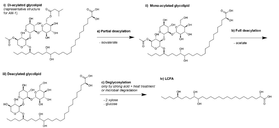
Based on previous in-vitro experiments in simulated gastric fluid and in pharmacokinetics studies in rats with [13C]-AM-1, it was hypothesized that AM-1 passes mostly unchanged to the lower GI tract after ingestion. Within the lower intestine, predominantly the ester linkages and partially the glycosidic linkages are prone to hydrolysis by microflora, to its components glucose, xylose, acetate, isovalerate and long chain fatty acids (LCFA) molecules, as shown in Figure 8.
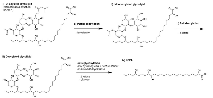
Since it was anticipated that its minor components, glucose, xylose, acetate, and isovalerate, would be further metabolized rapidly and incorporated for normal physiological functions, the major hydrolysis component of AM-1, i.e., LCFA, was studied in parallel to AM-1. This approach allowed the pharmacokinetics, excretion balance, and tissue distribution experiments to elucidate the ultimate fate of the parent compound, AM-1, by also studying its major hydrolysis product, LCFA.
For all study phases, rats received equimolar doses of either [14C]-AM-1 or [14C]-LCFA via oral or intravenous (IV) administration calculated on a mg/kg body weight basis using a dosage volume of 10 ml/kg for the oral route and 2 ml/kg for the IV route. This was followed by collection of biological samples at specified intervals. Details regarding the respective molecular weights (MW) used, dose groups, number of animals, and samples collected in each phase of the study are presented below.
For the repeated dose phases, rats received a single daily oral dose of either AM-1 or LCFA for 14 consecutive days followed by a single oral dose of [14C]-AM-1 or [14C]-LCFA. Following dosing in the pharmacokinetic phase, blood samples were collected from 3 animals/sex rotating among 3 sub-groups at selected time points through to 72 h post-dosing. In the excretion balance phases, expired air, activated carbon (from the activated carbon trap for volatile organic compounds), urine, faeces, cage rinse, cage wash, and select carcass samples were analysed for total radioactivity by liquid scintillation counting (LSC).
At approximately 1, 2, 4, 8, 24, 48, and 168 h post-dosing, 1 animal/sex/QWBA group was anesthetised with isoflurane and a whole blood sample of approximately 4 mL was collected via cardiac puncture into tubes containing K3EDTA as the anticoagulant. Concentrations of radioactivity were calculated in a range of selected tissues for both sexes.
Results
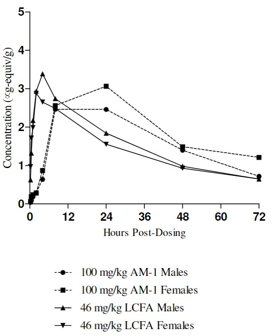
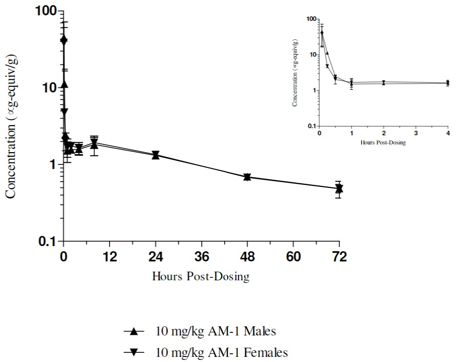
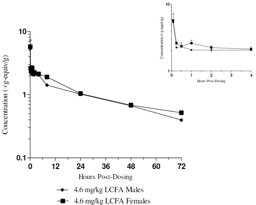
Circulating radioactivity at Cmax was equal to <0.1% (AM-1 equivalents) and 0.2% (LCFA equivalents) of the administered dose, if the approximate amount of circulating plasma in a 250g rat is assumed to be 7.8g. The initial phase in the pharmacokinetic (PK) profile, whose primary character is determined by absorption, differed between AM-1 and LCFA equivalents, Tmax was much earlier for LCFA equivalents (Figure 9).
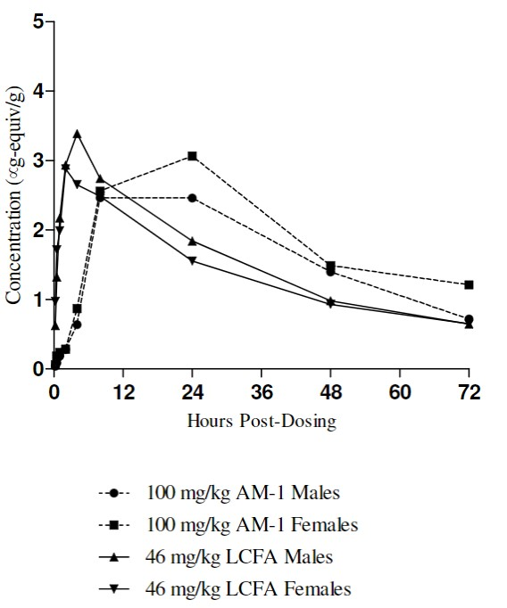
This was interpreted as indicating that orally administered LCFA was directly and rapidly absorbed, whereas AM-1 underwent several hydrolysis steps before its components were absorbed: therefore, accounting for the time delay in Tmax for AM-1. Furthermore, based on the kinetic profiles after intravenous administration, slow absorption of AM-1 followed by hydrolysis in the circulation may be occurring. There was approximately a 2-fold difference in the dose-normalized AUClast between AM-1 (approximately 1.3) and LCFA (approximately 2.3) equivalents, which is proportional to the higher molecular weight of AM-1. However, there was no apparent difference in the terminal elimination phase half-life between AM-1 and LCFA equivalents, which was determined to be approximately 30h.
Pharmacokinetic parameters for the [14C]-AM-1 and [14C]-LCFA IV-dose groups are presented in Table 23 and Table 24.
Higher C0 for AM-1 versus LCFA equivalents is tentatively attributed to a transient contribution from glucose, xylose, acetate, and isovalerate, following the hydrolysis of AM-1. Due to the short half-lives of these compounds, they are rapidly removed from circulating blood unlike LCFA, therefore resulting in the initially higher C0 for AM-1 compared with LCFA, followed by similar plasma concentrations for both substances.
There is approximately a 2-fold difference in the dose-normalized AUCinf between AM-1 (approximately 11) and LCFA (approximately 22) equivalents, which mirrors the two-fold difference in molecular weight of these compounds. Due to the similarity of the PK analysis, subsequent tissue distribution and the excretion balance behaviour, it was concluded that an equivalent concentration of LCFA (the ultimate hydrolysis product of AM-1) was present in the systemic circulation following equimolar administration of AM-1 and LCFA, suggesting that AM- 1 can be hydrolysed to LCFA in the circulation following an intravenous dose.
As the molecule is uniformly labelled, the calculations transforming administered radioactivity assume that test article equivalents result from AM-1 (average MW = 985) following administration of AM-1, when it is likely that there is a two-fold concentration of AM-1-derived LCFA (average MW = 455) with similar total mass.
Thus, as a function of moles of test article administered, the pharmacokinetic behaviour of AM-1 and LCFA equivalents following intravenous administration were virtually indistinguishable (Figure 10 and Figure 11).
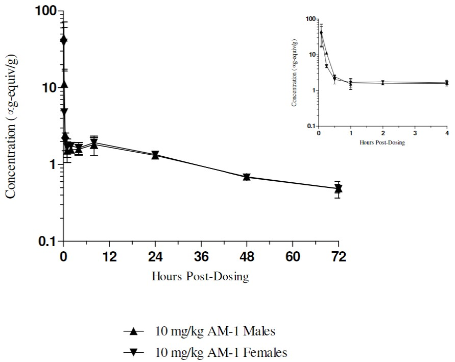
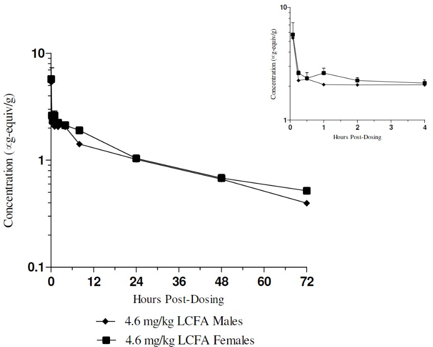
There was no apparent difference in the terminal elimination phase half-life between AM-1 and LCFA equivalents, which was determined to be approximately 37h (overall average). The similarity between AM-1 and LCFA equivalents in the terminal elimination phase following intravenous administration also indicates that AM-1 is at least partially hydrolysed in the circulation to LCFA.
Approximately 88% to 101% of the administered dose was recovered in expired air, urine, faeces, and carcass from male or female rats following single or repeated oral administration of [14C]-AM-1 at 100 mg/kg or single or repeated oral administration of an equimolar dose of [14C]-LCFA at 46 mg/kg. There appeared to be no difference in the excretion of AM-1 or LCFA equivalents based on sex (males versus females) or single versus repeated exposures. Approximately 15% to 21% of the recovered radioactivity was captured in the CO2 trap for expired air, approximately 1% to 3% of the recovered radioactivity was in the urine, and approximately 63% to 83% of the dose was eliminated in faeces. Possible sources of CO2 include intestinal metabolism of hydrolysis products by microbiota. It is likely that only a very small fraction of the AM-1 is hydrolysed before elimination: if a significant fraction were hydrolysed, then a strong contribution of the metabolites (other than LCFA) on the PK and excretion balance would be seen, especially differences in the percent of dose in the urine. Approximately 2% of the recovered radioactivity was in the carcass of the selected animals assessed. The majority of both test articles was eliminated in the faeces without absorption.
The apparent oral bioavailability of AM-1 and LCFA equivalents (approximately 11%; overall average for males and females combined) determined in the pharmacokinetic phase may reflect the absorption of degradation products of AM-1 and LCFA formed in the gut, including CO2 (which must be present systemically to be eliminated in the expired air), monosaccharides (from AM-1 only) and short chain fatty acids (SCFA). Quantitative analysis of the disposition of the radioactivity into the possible metabolic products formed prior to or after absorption is not practical due to the number of radiolabelled species and the number of absorption, distribution, and elimination processes occurring concurrently. However, the existing data support the conclusion that oral bioavailability of both AM-1 and LCFA equivalents is very low with ≤0.2% of the administered dose present in the plasma at Cmax, and less than 3.5% of the dose recovered in the urine.
[14C]-AM-1 or [14C]-LCFA derived radioactivity was detected by quantitative whole-body autoradiography in all tissues measured. Cmax and AUClast for [14C]-AM-1- and [14C]-LCFA derived radioactivity were highest in the tissues of the gastrointestinal tract, as expected following oral administration.
Eyes, testes, and brain showed only very low concentrations at a few time points in selected animals. The remaining tissues had concentrations of test article equivalents ranging from just above the LLOQ to <30,000 ng/g (excluding gastrointestinal tract), which represented a low concentration relative to the administered dose, and no target tissues were identified. Tmax was variable, depending on the tissue but generally correlated with the plasma or whole blood Tmax and was often earlier for LCFA than for AM-1.
For the majority of the tissues, T1/2 was considered not to be reportable or was an approximation; however, both AM-1 and LCFA concentrations were below the limit of quantification by 168h following either single or repeated dosing, suggesting tissue half-lives of approximately 24-40h, which is consistent with the observed plasma T1/2. Tissue: plasma ratios (calculated using AUClast) indicated that test article equivalents in tissue were primarily associated with the perfusion of tissues by whole blood and were < 0.7 for poorly perfused tissues. In addition to the similarity in tissue distribution between AM-1 and LCFA, there was no notable impact of single versus repeated dosing on the tissue distribution of either test article. Variations in the concentrations of AM-1 equivalents in tissues of the GI tract are expected based on individual variability in intestinal motility depending on the rat and when diet was ingested. The high concentrations in the various tissues of the GI tract, low concentration of circulating radioactivity, and low concentrations in the remaining tissues are consistent with low oral bioavailability, which suggests only a small portion of the administered AM-1 was absorbed into the systemic circulation, as was also evident in the autoradioluminograms.
Conclusion
The applicant concluded that AM-1 and LCFA and their gastrointestinal tract metabolites are poorly absorbed by the oral route and are primarily eliminated in the faeces without absorption. The pharmacokinetic, tissue distribution and excretion balance data derived are consistent with an interpretation that following ingestion, AM-1 is partially hydrolysed to its components, glucose, xylose, acetate, isovalerate and LCFA. The likely small primary metabolites glucose, xylose, acetate, and isovalerate are expected to have a fast absorption and high bioavailability but rapid clearance and thus expected to contribute marginally to the observed test article equivalents in blood and tissues after oral administration of AM-1. These results support an interpretation that systemic exposure to AM-1 or its metabolites would be very limited following oral ingestion. AM-1 is proposed for use as a food ingredient with antimicrobial / preservative properties without offering nutritional, taste or other technical benefits to foods or beverages. Therefore, limited absorption of AM-1 is a desirable property and serves to minimise exposure of the consumer to AM-1 or its major hydrolysis product, LCFA. In addition to limited exposure after a single dose, there was no change to the pharmacokinetics, distribution, and elimination after repeated exposures, which is positive regarding its application as a food ingredient where repeated ingestion is expected. Furthermore, because rapid hydrolysis and elimination of AM-1 occurs in vivo, the antimicrobial properties of the parent material would be absent or greatly diminished in the lower intestines so there is low concern for disruption of healthy gut microflora.
2.11.5. Antimicrobial properties of AM-1 in the gut
An analysis of the effects of AM-1 supplementation on the rat gut microbiome was submitted.
The study evaluates microbiome composition in rats following 90 days of exposure to AM-1 using 16S rRNA gene sequencing. The faecal and caecal samples analysed for microbiome effects were obtained from a 90-day OECD 408 toxicity study conducted on Sprague-Dawley rats. No adverse findings were identified in the study animals that would call into question the suitability of those animals for microbiome assessment, or the reliability of the resulting faecal and caecal microbiome data.
Doses tested were 333 mg/kg bw/day, 666 mg/kg bw/day and 1,000 mg/kg bw/day (highest dose), corresponding to approximately 523-, 1,046- and 1,570-fold higher exposures than the highest anticipated dietary exposure, respectively. Further details on Exposure estimates can be found in Section 2.10.
Faecal samples were collected on Days 44 and 90, and caecal contents collected on Day 91. A total of 120 samples were analysed using 16S rRNA amplicon sequencing to characterise the gut microbiota. Across the parameters assessed, there was little to no evidence of treatment-related effects on alpha diversity, beta diversity, or overall microbial community composition.
The study is not without limitations: it did not assess functional endpoints such as short-chain fatty acid production, microbial metabolism of AM-1, intestinal barrier integrity, absolute microbial abundance, or potential effects in human microbiota systems, but rather focusses on microbiome compositional analysis. In addition, the use of 16S sequencing does not provide strain-level resolution or direct information on microbial function. It is acknowledged that these limitations introduce uncertainty and mean that the study cannot fully address every theoretical concern that has been raised regarding potential microbiome effects.
However, the study did not identify any substantial perturbations of the gut microbial ecosystem, even at dose levels considerably higher than anticipated human dietary exposure. The results provide reassuring evidence that AM-1 does not produce significant adverse effects on the gut microbiota in this animal model.
Overall, the report findings indicate that on AM-1 supplementation for 90 days at doses up to 1,000 mg/kg bw/day, there is little to no detectable effect on microbiome diversity, microbiome structure, or taxonomic composition of the rat gut microbiota.
The FSA/FSS overall concluded that there are no significant concerns regarding the proposed uses of AM-1 of the gut microflora at the acceptable daily intake of 3.3mg/kg bw/day (details of the ADI can be found in section 2.13).
2.11.6. Genotoxicity
Genotoxicity of AM-1 was investigated by the Applicant in three studies: an in-vitro bacterial mutation assay (Ames test), an in-vitro human lymphocyte study (Micronucleus Test) and in-vitro mouse lymphoma assay.
In vitro bacterial mutation assay (Ames)
The Applicant conducted a study (TNO Triskelion, 2012) according to OECD TG 471 investigating the ability of AM-1 to induce reverse mutations in Salmonella typhimurium. This study was GLP compliant.
Five strains (TA98, TA100, TA1535, TA1537 and the Escherichia coli strain WP2uvrA) were exposed to AM-1 at five dose levels 62 – 5000 µg/plate. The plate-incorporation test with strain TA1537 was repeated at dose levels of 19, 38, 75, 150 and 300 µg/plate due to excessive cytotoxicity at dose levels ≥185 µg/plate in the first test.
Three bacterial reverse mutation tests were performed with dimethyl sulfoxide (DMSO) as a negative control, in the absence and presence of a metabolic activation system (S9-mix). After 48 to 72 hours of incubation at 37°C, the revertant colonies were scored.
The His+ and Trp+ revertants, respectively, were counted, toxicity was defined as a reduction by at least 50% in the number of revertant colonies and/or a clearing of the background lawn of bacterial growth as compared to the negative control and/or occurrence of pinpoint colonies.
In all tests, the mean numbers of His+ and Trp+ revertant colonies of the negative controls in all strains used were within the nominal acceptable range, except for the negative control for strain WP2 uvrA, in the presence of S9-mix, in the first test. The number of revertants was just above this range (62 instead of 60); this slight deviation was considered to be acceptable and not to influence the outcome of the study.
There were no test article-related increases in the mean number of revertant colonies compared to the background spontaneous reversion rate (negative control) in any strain tested either with or without the S9 mix.
It was concluded that the results obtained with the test article in Salmonella typhimurium strains TA1535, TA1537, TA98, TA100 and Escherichia Coli strain WP2uvrA, in both the absence and presence of the S9 mix indicate that AM-1 is not a base pair substitution or frameshift mutagen under the experimental conditions employed.
Full test conditions and study reports were supplied to the AEJEG and found to be acceptable.
In-vitro human lymphocyte study (Micronucleus Test)
The Applicant conducted a study (TNO Triskelion, 2012) according to OECD TG 487 investigating the potential of AM-1 to induce micronuclei in binucleated cultured human lymphocytes, in both the absence and presence of a metabolic activation system (S9-mix). This study was GLP compliant.
After a preliminary dose range finding cytotoxicity test, AM-1 was dissolved in DMSO and tested at concentrations of up to 700 and 1000 µg/mL in both the absence and presence of S9 for the 4 hour (pulse treatment) incubation. In the 20-hour (continuous treatment) incubation assay without metabolic activation, AM-1 was tested at concentrations up to 900 µg/ml.
For the main in vitro micronucleus test, both the 4-hour (pulse) incubation and the 20-hour (continuous) treatment groups were performed simultaneously; duplicate cultures were used.
After 48 hours, cells were harvested by low-speed centrifugation and resuspended in fresh medium, the test article (or control) was added. Incubations were continued for 4 hours (pulse treatment) or for 20 hours (continuous treatment). After incubation the culture medium was removed, the cells were washed twice before incubation for an additional 20 hours (pulse treatment) and 28 hours (continuous treatment) prior to being harvested (second cell-cycle). Based on the evaluation of cytotoxicity in all treatment groups (±S9 pulse and-S9 continuous), three dose levels of the test article together with the negative control and positive control, were analysed for micronucleus induction in bi-nucleated lymphocytes. At the end of the recovery period the cells in each culture were harvested and processed. Two thousand (2000) binucleated cells per concentration (1000 per culture) were examined for the presence of micronuclei.
The dose levels selected for the treatment with and without metabolic activation have been provided in Table 26.
Outcomes of the negative and positive control demonstrated all criteria for a valid test were met. Dose related cytotoxicity was observed in both pulse treatment (± S9) and continuous treatment (-S9) groups. Furthermore, the highest concentrations selected for micronuclei analysis were within the testing guidelines specified range of 55 ±5% cytotoxicity.
The Applicant concluded that under the experimental conditions employed, AM-1 was not clastogenic and / or aneugenic to cultured human lymphocytes.
Full test conditions and the study report were supplied to the AEJEG and were found to be acceptable.
In vitro mouse lymphoma assay
The Applicant conducted a study (WIL Research, 2016) which was OECD TG 490 compliant. The study was GLP compliant except for the test item being research grade and that the analytical characterisation conducted by the supplier was non-GLP, yet the COA for the batch was generated using a validated method and established under standard operating procedures.
The study was performed to evaluate the mutagenic activity of AM-1 in an in-vitro mammalian cell gene mutation test with L5178Y/TK +/- mouse lymphoma cells, in both the absence and presence of a metabolic activation system (S9-mix).
After a preliminary dose range finding cytotoxicity test, AM-1 was dissolved in the solvent DMSO and tested at concentrations of up to 450 and 800 µg/mL in the presence and absence of S9 for the 3-hour respectively incubation. In the 24-hour incubation assay without metabolic activation, AM-1 was tested at concentrations up to 500 µg/ml. The highest doses that were tested gave a cell survival of approximately 10-20% and survival at the lowest doses was approximately the same as of the cells in the solvent control.
Methyl methanesulfonate (MMS) was used as a direct acting mutagen and cyclophosphamide (CP) was used as an indirect acting mutagen as positive controls.
In the main study cultures (106 cells/ml for 3-hour treatment and 1.25x 10⁵ cells/ml for 24 hour treatment) were incubated with either AM-1, negative or positive controls. All incubations were carried out in a humid atmosphere (42-90%) containing 5 ± 0.5% CO2 in the air in the dark. After exposure cells were separated from treatment solutions by centrifugation, cells in the final suspension were counted with a coulter particle counter, remaining cells were cultured for two days after the treatment period for expression of the mutant phenotype.
Two days after the end of the treatment with the test article the cells were plated for determination of the cloning efficiency (CEday2) and the mutation frequency (MF). These were then incubated for 10–12 days, stained and scored with the naked eye or by light microscopy.
In the absence of S9 mix, AM-1 did not induce a significant increase in the mutation frequency in the 3-hour exposure, which was then confirmed in an independent experiment with modification in the duration of treatment (24-hour exposure). In the presence of S9 in the 3-hour exposure AM-1 did not induce a significant increase in the mutation frequency. Results for both positive and negative controls demonstrated all criteria for a valid test were met.
The Applicant concluded that AM-1 is not mutagenic in the TK mutation test system, both with and without metabolic activation under the experimental conditions employed.
Full test conditions and the study reported were supplied to the AEJEG and found to be acceptable.
2.11.7. Toxicity testing (subchronic, chronic and carcinogenicity)
Subchronic (Repeated Dose) Toxicity Studies with AM-1
90-Day Oral (Drinking Water Administration) Toxicity Study in Rats
The Applicant conducted a 90-day oral toxicity study (drinking water administration) (LPT, 2015) according to OECD TG 408 in male and female CD Crl:CD(SD) rats. This study was GLP compliant.
A preliminary dose range-finding toxicity study with a maximum dose level of 1% AM-1 tested in drinking water for 14 days was performed no test article-related signs of systemic toxicity were noted. Following this preliminary dose, range-finding study a 90 day study was undertaken in which groups of animals were orally administered the vehicle control and test article (AM-1) diluted with tap water at concentrations of 0%, 0.15%, 0.5% and 1.5% (0, 1.5, 5.0 and 15mg/mL respectively) for 90 days.
A standard listing of clinical chemistry and urinalysis parameters were analysed in accordance with the applicable testing guidelines of OECD 408 and FDA Redbook.
On Day 91 (main study, one day after the last administration of the test article) or Day 119 (recovery phase, 28 days following cessation of test article administration), a complete gross necropsy was performed on all animals euthanised under CO2 anaesthesia and exsanguinated. The organs and tissues of all main study animals of the control and high-dose (1.5% AM-1) treatment groups and any prematurely deceased animals were examined microscopically after preparation of paraffin sections and haematoxylin and eosin (H&E) staining. The stomachs of all main study animals in the low- and mid-dose (0.15% and 0.5%) treatment groups as well as all recovery animals were examined histologically due to observations in the stomach/forestomach of the main study animals in the high-dose (1.5% AM-1) group. Additional microscopic examinations were performed on the heart, liver, and one kidney (males and females using Oil Red O staining) and on one testis and one epididymis (males only using periodic acid-Schiff (PAS) staining) from the main study animals in the control and high-dose (1.5% AM-1) treatment groups.
One of 30 female rats treated with 1.5% AM-1 via the drinking water died during test day 14 after blood withdrawal for laboratory examinations. The death of the animal was regarded as incidental and related to the stress caused by the isoflurane anaesthesia during the blood withdrawal procedure. The animal was in a poor physical condition due to fairly low food and drinking water consumption during the first test week. The death of the animal was regarded to be a secondary effect and not related to the treatment with the test item, as no premortem behavioural changes were noted. Neither the macroscopic inspection at necropsy nor the histopathological examination revealed any test item-related organ changes that could be correlated with the premature death of the animal. None of the male animals of the high dose group treated with 1.5% AM-1 via the drinking water nor any other female rats of the high dose group died prematurely during the treatment period.
A decrease in drinking water consumption was noted for male and female rats treated with 1.5% AM-1 via drinking water, particularly in weeks 1 and 2. The drinking water consumption of the rats treated with 0.5% or 1.5% AM-1 decreased throughout the treatment period in a dose-dependent manner, which is most likely due to a fairly high viscosity and/or a decreased palatability of the drinking water containing 0.5% or 1.5% AM-1.
The food consumption of both male and female rats treated with 1.5% AM-1 was decreased in study week 1, and the body weight of the 1.5% dose male animals was marginally below the body weight of the control animals from study week 1 and throughout the treatment period. The differences detected were only statistically significant during the first 6 weeks of the study, female bodyweights for the 1.5% dose were unaffected.
It is well known in the scientific literature that under normal circumstances rats maintain a fairly constant ratio of food intake to water intake. Thus, reduced water intake is associated with reduced food intake and a reduction in body weight gain. This voluntary reduction in food intake and thus growth supports the constant ratio of body weight to lean body mass (Collier & Knarr, 1966; Crampton & Lloyd, 1954; Jackson & Smith, 1931).
The statistically significant differences in relative food consumption compared to the control group are not considered to be test item-related, as depicted in Table 27.
The body weight, body weight gain and body weight at autopsy were not affected in the male and female animals treated with 0.15% or 0.5% AM-1 and in the female animals treated with 1.5% AM-1. The body weight of the male animals treated with 1.5% AM-1 was marginally reduced (statistically significant at p ≤ 0.01 or p ≤ 0.05) in test weeks 1 to 6 (test days 8, 15, 22, 29, 36 and 43) by 5-7% compared to the control group. This decrease in body weight corresponds to the initial decreased food and drinking water intake during the first one or two test weeks. Although the food and drinking water intake of the rats normalised as of test weeks 2 (food consumption) and 3 (water consumption) onwards, respectively, the body weight of the male high dose animals remained marginally below the body weight of the control group animals (less than 5%) throughout the treatment period.
At autopsy body weight of the male animals treated with 1.5% AM-1 via drinking water was reduced by 2.1% compared to the control group (no statistical significance).
In conclusion, the slightly reduced body weight of the male animals treated with 1.5% AM-1 is considered to be due to the reduced food and drinking water intake of the rats and not due to a systemic toxic effect of the test item. This is supported by the fact that the body weight of these animals almost completely recovered compared to the control group during the study.
In contrast to the effects reported in this drinking water study, repeated oral administration of AM-1 via gelatine capsule administration at up to 1000 mg/kg bw per day was well tolerated in Beagle dogs (study summarized below) and only minimal differences in food consumption (not statistically significant) and cumulative body weight gain were noted in the high-dose (1000 mg AM-1/kg bw per day) females. Therefore, the differences observed in the present drinking water study were considered to be an artefact of the route of dose administration related to high viscosity, surfactancy, and reduced palatability, and not an indication of systemic toxicity of the test article.
The statistically significant differences noted for selected clinical pathology parameters such as Erythrocytes (RBC), Hematocrit (%), MCV (fL), MCH, Albumin, Globulin, Protein, Calcium, Sodium, Chloride, ALAT, and LDH were regarded as secondary stress-related effects due to decreased drinking water intake and the resulting slight dehydration of the animals and not due to a direct systemic toxic effect of the test article. Consistent patterns that would suggest biological significance were not observed for several statistically significant parameters, since the results were within the laboratory’s historical control data ranges, were not dose-related, and/or lacked correlation with histopathological findings.
No adverse test article related effects were noted on haematological and serum chemistry parameters, urinalysis, eyes or optic region, relative body weight at necropsy and absolute organ weights at any dose levels at the end of the treatment period. There were no test article related changes upon macroscopic inspection, microscopic examination of all scheduled organs and tissues of the 1.5% dose, gross lesions of the 1.5% dose animals and the stomachs of the 0.15% and 0.5% dose rats did not reveal any morphological changes that are considered to be related to the administration of the test article.
A very small number of macroscopic changes were noted in the kidney (tissue enlargement), thymus (red discolouration), uterus (dilated, filled with clear liquid), ovaries (cystic) and eyes (reduced in size) of individual animals of both the test item-treated groups and the control group at termination at the end of the treatment period and the recovery period. These changes are considered to be incidental findings due to the low number of animals affected and lack of a dose-response relationship.
The tissue enlargement of the right kidney noted in 1 of 20 male animals treated with 1.5% AM-1 via the drinking water at termination correlated with a benign nephroblastoma. However, this finding was considered to be incidental and without any toxicological relevance as only one animal of the high dose group was affected.
The gross lesions of groups 2 and 3 (females: group 2: 1/20 animals: ovaries cystic, 2/20 animals: uterus dilated, filled with clear liquid; females: group 3: 2/20 animals: uterus dilated, filled with clear liquid), except stomachs, were not examined.
No test item-related effects were noted on the relative (to body weight at necropsy) and absolute organ weights of the male and female animals treated with 0.15%, 0.5% or 1.5% AM-1 via the drinking water compared to the control group, either at termination at the end of the treatment period, or at the end of the recovery period.
The statistically significant (at p ≤ 0.05 or p ≤ 0.01) increase observed for the relative and absolute stomach weights in the intermediate and high dosed female animals compared to the control group at the end of the treatment period is regarded to be without any toxicological relevance, as they were not clearly dose-related, not consistent between sexes and no histopathological findings were noted which could be correlated to this finding.
Statistically significant differences in absolute or relative organ weights at scheduled termination on test day 91 which were not considered to be test item-related are given in Table 28 and Table 29.
Statistically significant differences in absolute or relative organ weights at recovery termination on test day 119, which were not considered to be test item-related, are given in Table 30 and Table 31.
At the end of the 28-day recovery period the mean body weights for the 1.5% dose were within the range of the control group, supporting the conclusion that the slightly decreased body weights of the 1.5% dose observed during treatment were associated with reduced intake of the treated drinking water.
Clinical parameters in the 1.5% dose groups animals were comparable to control group values at the end of the 28-day recovery period with the exception of certain parameters in males (Red Blood Cells (RBC), Large Unstained Cells (LUC), Mean Corpuscula Volume (MCV), Mean Corpuscular Haemoglobin (MCH), plasma albumin and calcium levels and LUC and thromboplastin time (TPT) in females. Changes in these parameters appear to be spurious statistical variations and without biological relevance due to only slight differences from controls, some occurred due to the control values being on the high or low end of the historical control range.
The Applicant concluded minor variations in some parameters such as body weight and select clinical chemistry were evaluated and considered to be incidental and secondary to the observed reduced drinking water consumption / mild dehydration due to the reduced palatability of the test article treated drink water.
Therefore, the no observed adverse effect level (NOAEL) for systemic toxicity was considered to be 1.5% AM-1 in drinking water, equivalent to 1201 and 1423 mg/kg bw per day for male and female rats, respectively. This NOAEL is corroborated by the results of a 90-day subchronic oral (capsule administration) toxicity study of AM-1 in Beagle dogs in which no adverse effects were observed at doses up to 1000 mg/kg bw per day.
The AEJEG were satisfied there was no toxicity concern and the NOAEL of 1000 mg/kg bw per day was agreed by the AEJEG.
90-Day Oral (Capsule Administration) Study in dog
The Applicant conducted a subchronic toxicity study (2016) according to OECD guideline 409 evaluating AM-1 in a 90-day oral study in male and female Beagle dogs. The study was GLP compliant except for the characterisation analysis of the test item conducted by the supplier, this was not conducted to GLP or GMP standards but conducted according to the suppliers established standard procedures.
Oral administration was chosen as this is the intended route of exposure, the move to using a capsule to administer the test substance was to mitigate the palatability issues observed in the rat 90-day study (LPT, 2015).
The test article was administered by oral capsule at doses of 150, 500, 1000 mg/kg bw per day for 90 days, with control group animals receiving the same number of empty capsules as the high dose (1000mg/kg bw per day) group. Each animal received an approximate 5 – 15mL flush of water using a graduated syringe following administration.
A standard listing of clinical chemistry and urinalysis parameters were analysed in accordance with the applicable testing guidelines of OECD and FDA Redbook. A complete gross necropsy was performed on all animals, the organs and tissues of all animals of the control and high-dose treatment groups were examined microscopically after preparation of paraffin sections and haematoxylin and eosin (H&E) staining. Gross lesions were examined microscopically from all animals.
There were no test article-related deaths or changes in behaviour or external appearance of the study animals, however there were clinical observations attributed to the capsule administration method including emesis, clear or frothy (white) material around the mouth and salivation. Abnormal (soft or mucoid) faeces and diarrhoea were noted occasionally and occurred in similar incidence in the control groups; therefore, these observations were not considered test article related. A single occurrence of tonic convulsions was noted in 150mg/kg/day group Male (ID number 9921) on Study Day 88. This observation was a single occurrence in the low-dose group, and it was not likely to be related to test substance administration.
A minimal test article related reduction in body weight compared to the control group, determined to be not statistically significant at any weekly interval, was observed in the 1000 mg AM-1/kg bw per day group for females; this was approximately 6% by study week 12. The cumulative body weight gains in the 1000mg AM-1/kg bw per day group for females were statistically significantly lower than the control group, and a minimal reduction in food consumption was also observed, although when compared to the control group this was not statistically significant.
The AEJEG commented how the body weight change occurred in the week one female group, however the same group showed a weight gain in the following weeks. Members agreed that the effect noted was likely to be a palatability issue.
It was speculated that AM-1, which has surfactant properties at high aqueous concentrations, may have made the dogs feel unwell for some time after dosing because of high concentrations in the stomach contents after release from administered capsules, resulting in slightly lessened appetite. No other test article-related effects on food consumption or body weights were noted.
Statistically significant changes were noted infrequently for some haematological, coagulation, or serum chemistry parameters; however, these differences were not considered test article related as they were minimal and/or lacked a dose response, or values were similar to those measured during acclimation or were within the laboratories’ historical control ranges. Additionally, inconsistent statistically significant findings may also be related to random variation seen in studies with small group sizes. No test article-related effects were noted on the urinalysis parameters. The Applicant concluded there were no gross necropsy observations that were considered to be associated with the administration of the test substance.
Higher epididymides weight relative to final bodyweight was noted in the 150 and 1000 mg/kg/day group males, higher heart weight relative to final body weight was noted in the 150 mg/kg/day group females, and higher liver weight relative to final body weight was noted in the 150 and 1000 mg/kg/day group females. All differences noted in organ weights between the control group and the treated groups were not considered test substance related due to the lack of a dose dependent relationship.
The microscopic examination of all scheduled organs and tissues of the 1000mg AM-1/kg bw per day group for male and female animals and gross lesions from all animals showed no signs of test article-related histologic alterations. Histological changes were considered to be incidental findings or were related to some aspect of the experimental manipulation other than administration of the test article.
One 1000 mg/kg/day group female had portal vein hypoplasia, yet this finding was considered to be a congenital alteration and not related to the test substance. There were no test substance-related changes in the prevalence, severity, or histologic character of those incidental tissue alterations.
In conclusion, oral administration of AM-1 to sexually mature Beagle dogs at dose levels of 150, 500, and 1000 mg/kg bw per day for a minimum of 91 days was well tolerated at all dosages. Test substance-related changes were limited to minimal effects on food consumption and body weights in the 1000 mg/kg bw per day group females, however, mean body weights for the high dose group females were not statistically significantly different from controls during any weekly interval of the study.
The NOAEL was considered to be 1000 mg/kg bw per day, which was the highest dosage level tested. This NOAEL is corroborated by the results of the 90-day oral (drinking water administration) toxicity study in rats (i.e. 15 mg/mL in drinking water, equivalent to 1201 and 1423 mg AM-1/kg bw per day for male and female rats, respectively), in which observed differences in drinking water consumption, body weights, and clinical pathology parameters were considered incidental to the route of dose administration and viscosity of the drinking water containing higher concentrations of AM-1.
The AEJEG noted that whilst the original study report concluded the same NOAEL figures as above, the study also concluded that the no-observed-effect level (NOEL) was considered to be 1000mg/kg/day for males and 500mg/kg/day for females.
Graphical representation of endpoints for which statistically significant differences were observed but argued away was later provided by the Applicant, in addition to comparisons of these graphs with historical controls.
The AEJEG was not satisfied with the information provided on the 90-day dog study and commented that they did not agree with FSANZ that there was a clear effect from this study. The AEJEG concluded that there was little toxicity noted. The AEJEG also highlighted that this was not a long-term study and that the low number of animals used meant that the results could not be considered statistically significant.
The AEJEG were more satisfied with the rat study for determining the NOAEL. This was due to the study design having more animals in each test group in comparison to the dog study, giving more statistical significance to the results. Members also agreed the rat study was more comparable with how humans would consume the product as this was added to the rats’ drinking water, as opposed to taken in the form of a capsule in the dog.
Chronic toxicity and carcinogenicity
Assessment of Mutagenicity and Carcinogenicity Potential for AM-1
The Applicant stated based on the chemical structural characteristics of AM-1, (glycolipid mixture with no reactive groups), there was low potential for carcinogenicity or mutagenicity from dietary intake of this material. The metabolism profile for AM-1 was well understood and allowed the conclusion that there are no carcinogenic or mutagenic metabolites of AM-1 formed in vivo. No target organs were identified and no accumulation were found in the ADME study. Finally, genotoxic studies did not indicate any risk for mutagenic (or carcinogenic) effects.
Chronic toxicity of AM-1
The Applicant stated, based on the results of the performed Tier-1- and Tier-2 testing as laid out in the EFSA guidance (EFSA, 2012) (ADME, genotoxicity, acute toxicity, irritation and sensitization studies, subchronic toxicity, reproductive and developmental toxicity), there was a lack of systemic availability demonstrated and an absence of genotoxicity in vitro, none of the triggers for justifying the need for a chronic toxicity study were activated.
Assessment of Mutagenicity and Carcinogenicity Potential for AM-1
The Applicant stated based on the chemical structural characteristics of AM-1, (glycolipid mixture with no reactive groups), there was low potential for carcinogenicity or mutagenicity from dietary intake of this material. The metabolism profile for AM-1 was well understood and allowed the conclusion that there are no carcinogenic or mutagenic metabolites of AM-1 formed in vivo. No target organs were identified and no accumulation was found in the ADME study. Finally, genotoxic studies did not indicate any risk for mutagenic (or carcinogenic) effects.
Chronic toxicity of AM-1
The Applicant stated, based on the results of the performed Tier-1- and Tier-2 testing as laid out in the EFSA guidance (EFSA, 2012) (ADME, genotoxicity, acute toxicity, irritation and sensitization studies, subchronic toxicity, reproductive and developmental toxicity), there was a lack of systemic availability demonstrated and an absence of genotoxicity in vitro, none of the triggers for justifying the need for a chronic toxicity study were activated.
2.11.8. Reproductive and developmental toxicity
Rat Two-Generation Reproduction Toxicity Study with AM-1
The Applicant conducted a Two-Generation Reproduction Toxicity study in Crl:CD(SD) rats (2016) according to OECD TG 416. The study was GLP compliant except for the characterisation analysis of the test item conducted by the supplier which was not conducted to GLP or GMP standards but conducted according to the suppliers established standard procedures.
The study consisted of 3 test article treated groups and one control group, composed of 25 rats/sex/group. AM-1 was administered daily by oral gavage for at least 70 consecutive days prior to mating. Dosing levels were 150, 500 and 1000 mg/kg bw per day. F0 animals were 6 weeks old at the initiation of test article administration, the test article was administered to offspring selected to become the F1 parental generation following weaning i.e., beginning on post-natal day (PND) 21.
The F0 and F1 males continued to receive the test article throughout mating and through the day prior to euthanasia. The F0 and F1 females continued to receive the test article throughout mating, gestation, and lactation, and through the day prior to euthanasia. Offspring (25/sex/group) from the pairing of the F0 animals were selected by PND 21 to constitute the F1 generation. Non-selected F1 pups and all surviving F2 pups were euthanized on PND 21. F0 males and females were dosed for 128–133 consecutive days, and F1 males and females were dosed for 138–148 consecutive days.
Individual F0 and F1 male body weights were recorded weekly throughout the study and prior to the scheduled necropsy. Once evidence of mating was observed, female body weights were recorded on Gestation Days 0, 4, 7, 11, 14, 17, and 20 and on Lactation Days 0 (when possible), 1, 4, 7, 11, 14, 17, and 21.
F0 and F1 male and female food consumption was measured weekly except during cohabitation. For females with evidence of a positive mating, food consumption was measured during gestation and lactation at the same intervals as body weight measurements.
Vaginal lavages were performed daily and evaluated microscopically to determine the stage of the oestrous cycle of each F0 and F1 female for 21 days prior to cohabitation and continuing until evidence of mating was observed or until the end of the mating period.
The F0 and F1 animals were paired on a 1 male to 1 female basis within each treatment group after a minimum of 70 days of treatment. Positive evidence of mating was confirmed and was defined as Gestation Day 0. All females were allowed to deliver naturally, and the day of parturition was designated PND 0. Beginning on PND 0, pups were sexed and examined for gross malformations, and the numbers of stillborn and live pups were recorded. Individual gestation length and mating, fertility, copulation, and conception indices were calculated.
A minimum of 1 male and 1 female F1 pup/litter from each treatment group were randomly selected prior to weaning (PND 21) to comprise the F1 generation (25 rats/sex/group).
Following the completion of weaning of the F1 and F2 offspring, all surviving F0 and F1 adults received a detailed physical examination, females received a vaginal lavage to determine state of the oestrous cycle and all were euthanised.
Spermatogenic endpoints (sperm motility [including progressive motility], morphology, and numbers) were recorded for all F0 and F1 males, and ovarian primordial follicle counts were recorded for all F1 females in the control and high dose groups and for all F1 females suspected of reduced fertility. For females that delivered or had macroscopic evidence of implantation, the numbers of former implantation sites were recorded. The number of unaccounted-for sites was calculated and the numbers of corpora lutea and former implantation sites were also recorded for females necropsied during gestation through Lactation Day 4.
The testing guideline-required list of tissues and organs were weighed and/or retained from all parental F0 and F1 rats. Microscopic evaluations were performed on select tissues for all parental F0 and F1 animals in the control and high-dose groups and for all animals found dead. Reproductive organs of the F0 and F1 adult rats suspected of reduced fertility were examined microscopically. Gross lesions were examined from all animals in all groups.
A few non-test article-related deaths occurred in the F0 and F1 generations which were considered the likely result of the intubation error based on macroscopic findings noted at necropsy (including dark red discoloration and/or areas of the lungs, lungs that were not fully collapsed, and/or foamy tracheal contents) and microscopic finding of inflammation, haemorrhage, and/or oedema of the lungs in multiple animals that may have been related to inadvertent pulmonary aspiration of the test article during or following gavage.
Test article-related increased incidences of rales and red and/or clear material around the nose and/or mouth were noted in the 150, 500, and 1000 mg/kg/day group F0 and F1 males and females compared to the control group occasionally at the detailed physical examinations or daily examinations, and more frequently at 1-2 hours following dose administration. These findings were generally noted in a dose-related manner throughout the treatment period for both generations. Red and/or clear material findings around the nose and/or mouth are common following oral gavage administration and although dose-responsive, these observations only occasionally persisted to the detailed physical examinations or daily examinations, and therefore were considered non-adverse in the absence of other signs of systemic toxicity. The increased occurrence of rales 1-2 hours following dose administration was attributed to the surfactant properties of the test article combined with oral gavage dosing and was not considered specific to the test article. Similar observations of respiratory distress related to aspiration of an irritant dosing material, especially following treatment with the more concentrated/viscous suspensions at higher doses, were reported in rat and rabbit developmental toxicity studies conducted with ethyl lauroyl arginate (i.e. LAE) via gavage (EFSA, 2007).
No test article-related effects on mean body weights and body weight gains (including body weights during gestation and lactation) were noted in the 150, 500, and 1000 mg/kg/day group F0 males and females or the F1 females. The values in the test article-treated groups were generally similar to the control group values for the pre-mating period (females) or the entire generation (males).
Significantly (p < 0.05 or p < 0.01) lower mean body weight gains were transiently observed in the 150 mg/kg/day group F0 males during Study Days 28–35 and the 500 mg/kg/day group F0 males during Study Days 0–7, 28–35, and 70–77. In addition, a significantly (p < 0.01) higher mean body weight gain was noted in the 150mg/kg/day group F0 males during Study Days 63–70. A significant (p< 0.01) mean body weight loss was noted in the 1000mg/kg/day group F0 females during Study Days 63-70. These were not attributed to test article administration because they were transient, did not occur in a dose-related manner, and/or had no effect on mean absolute body weights. Other differences from the control group were slight, did not occur in a dose-related manner, and/or were not statistically significant.
Mean maternal body weights and body weight gains were unaffected by test article administration during gestation. Differences between the control, 150, 500, and 1000mg/kg/day groups were slight and not statistically significant apart from a significantly (p < 0.01) lower mean body weight gain noted in the 1000mg/kg/day group compared to the control group during Gestation Days 11–14, Figure 12.
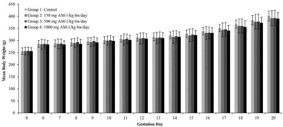
Mean maternal body weights and body weight gains were unaffected by test article administration during lactation. Differences between the control, 150, 500, and 1000mg/kg/day groups were slight and not statistically significant apart from a significantly (p < 0.05) lower mean body weight gain which was noted in the 500mg/kg day group when compared to the control group during Lactation Days 1–4. The transient reduced mean body weight gain for both exceptions to gestation and lactation given above did not affect mean absolute body weights or the overall body weight gain in these groups, and therefore was not attributed to test article administration.
Test article-related lower mean body weight gains were noted in the 150, 500, and 1000 mg/kg/day group F1 males during PND 21–28, when pups were just weaned and first receiving oral gavage doses of the test article, the differences from the control group were significant (p < 0.05 or p < 0.01).
Mean body weight gains were generally similar to the control group throughout the remainder of the study, with the exception of a few significantly (p < 0.05 or p < 0.01) lower body weight gains that were transient and/or not dose related. Mean body weight gains in the 150, 500, and 1000mg/kg/day group males were lower (not statistically significant) than the control group when the overall pre-mating period (PND 21–98) and entire generation (PND 21-161) were evaluated. As a result of the initial lower mean body weight gains during PND 21 28, mean body weights for F1 males were between 4.0% and 7.1% lower than the control group throughout the study (Figure 13). These differences were not considered adverse because the mean body weights in these groups at termination (PND 161) were only 5.4% to 6.1% lower than the control group, demonstrating that the initial effects were ameliorated over the course of the generation.
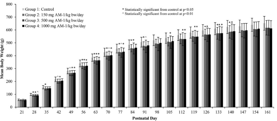
Mean food consumption evaluated as g/animal/day, and food efficiency in the 150, 500, and 1000 mg/kg/day groups for the F0 and F1 males and females was unaffected by test article administration throughout the study (including gestation and lactation). Within the 1000mg/kg/day F1 males group a significantly (p < 0.05) lower mean food efficiency value was noted during the last week of the study. However, mean food consumption and body weight gain in this group were comparable to the control group during this interval, and therefore the lower mean food efficiency was not considered test article-related.
No test article-related effects on F0 and F1 reproductive performance, including mating and fertility, male copulation and female conception indices, oestrous cycle lengths, pre-coital intervals, gestation length, the process of parturition and spermatogenesis parameters (motility, progressive motility, testicular and epididymal sperm concentration, sperm production rate, and the percentage of morphologically normal sperm), were observed at any dosage level. In addition, there were no test article-related macroscopic or microscopic findings or effects on organ weights noted for F0 and F1 males and females at any dosage level, or on the F1 female primordial follicle counts.
The mean number of pups born, live litter size, percentage of males per litter at birth, and postnatal survival through PND 21 were unaffected by administration of the test article to the F0 and F1 parental animals at all dosage levels. The F1 and F2 pups did not display any effects of test article exposure as evaluated by their general physical condition, body weights, necropsy findings, or organ weights.
Based on the lack of effects on F0 and F1 reproductive performance (mating, fertility, copulation and conception indices, oestrous cyclicity, and spermatogenic endpoints), the no-observed-adverse-effect level (NOAEL) for parental reproductive toxicity of AM-1 when administration orally via gavage to Crl:CD(SD) rats was 1000 mg/kg/day. There were no adverse effects on survival, clinical observations, body weight or food consumption parameters, macroscopic or microscopic findings, or organ weights for F0 or F1 males and females at any dose level.
Based on these results, a dosage level of 1000 mg/kg/day was considered to be the NOAEL for F0 and F1 parental systemic toxicity. There were no test article-related effects on F1 and F2 postnatal survival, development, or growth during the pre-weaning period, and therefore, the NOAEL for neonatal toxicity was considered to be 1000 mg/kg/day.
The AEJEG commented that some observed effects or statistically significant differences had been disregarded without clear reason. The AEJEG requested the Applicant to provide clear reasoning on the justification of why effects and statistically significant differences have been disregarded.
In response the Applicant provided clarification to the AEJEG on the endpoints such as food intakes and body weights for all generations for which some of the statistically significant effects and changes had been disregarded. Other endpoints the AEJEG requested clarification on were PND 0 Litter Data and Postnatal Survival, Organ Weights, F1 male and female food consumption and food efficiency and F2 PND 0 Litter Data and Postnatal Survival.
The AEJEG found the explanation acceptable and agreed with the conclusions of the study report.
2.11.9. Other Toxicity
Allergenicity
The Applicant provided the allergenicity claims and considerations as presented below.
Glycolipids were not identified as food allergens or mediators of food allergy in comprehensive scientific opinions by authoritative bodies (e.g. (EFSA, 2014; NIAID, 2011)). AM-1 does not contain,nor does the equipment used to process AM-1 contain, any of the food allergens listed in Annex II of the Regulation (EC) 1169/2011 of Food Information to Consumers.
AM-1 may contain up to 3% total protein content, which is associated with non-viable cell debris originating from the production organism, Dacryopinax spathularia, but does not contain any other introduced protein that would be expected to result in an allergic reaction.
Exposure to red mammalian meat containing the oligosaccharide galactose-α-1,3-galactose (α-Gal) expressed in glycoproteins or glycolipids has previously caused allergic reactions among individuals who have become sensitized to α-Gal after tick bites (Steinke et al., 2015; Wolver et al., 2012), however, α-Gal or similar carbohydrates are not present in the AM-1 glycolipid mixture.
To the best knowledge of the Applicant, food types listed as major food allergens do not contain glycolipids similar to AM-1.
The presence of glycolipid type compounds such as sphingomyelin, which belong to the class of sphingolipids has been reported in food such as milk, eggs, and soybeans (Fujino et al., 1970; Jyonouchi et al., 2011; Leray, 2015; Newberg & Chaturvedi, 1992).
The ability for milk-derived sphingomyelin to engage invariant natural killer T cells (iNKTs) and T-cell receptors (TCRs) has been reported, although the role of iNKTs in food allergy is still not fully understood (Jyonouchi et al., 2011). However, the Applicant claims these molecules are very different in their chemical structures and molecular properties compared to AM-1.
A single investigation regarding the potential for peanut glycolipid antigens to activate immune mechanisms related to food allergy was identified in the NIH Research Portfolio Online Reporting Tool (RePORT) (Umetsu, 2010); however, the results of this research have not been reported and the Applicant claims peanut “lipid and lipid-carbohydrate complexes described in the grant abstract are not considered to be similar to AM-1 glycolipids”.
Finally, AM-1 is not well absorbed via the oral route and there were no test article-related effects on any haematological parameters related to immune system responses in the repeat-dose oral toxicity studies in rats and dogs. However, the AEJEG noted that this was not a sufficient argument as effects may occur at very low doses and not be seen at very high doses.
AM-1 was also non-sensitising in dermal sensitisation studies in animals and humans summarised previously; however, the relevance of dermal sensitization models to distinguish allergenic from non-allergenic food extracts is still questionable (Selgrade, et al., 2009).
In consideration of the above information, it can be concluded that there is negligible potential for AM-1 to induce symptoms of food allergy.
The AEJEG considered the Applicant’s conclusion satisfactory.
Additional toxicity testing
The Applicant also provided the below toxicity studies, which have been summarised with the conclusion in Table 32.
The AEJEG was satisfied with the studies provided and the conclusions made.
2.12. Risk Assessment of 3-hydroxy-3-methylglutarate (HMG)
2.12.1. Safety of HMG
The presence of glycolipids containing 3-hydroxy-3-methylglutarate (HMG) as an acyl unit has been noted in certain batches of AM-1, including those batches of AM-1 tested in the 90-day rat and dog toxicity studies, rat ADME study, and rat developmental and reproductive toxicity studies. HMG is a minor hydrolysis product of a minor component of AM-1 with a calculated average concentration of <1.5% (w/w).
It was noted that the Applicant calculated an average concentration of <1.5% (w/w) within their Technical Dossier however the Applicant also provided documentation in which they calculate the theoretical maximum amount of HMG obtained from AM-1 to be <2.52% (w/w).
The maximum ratio of HMG-bearing glycolipids is fixed as <17% by the internal specification of AM-1 (verified analytically by HPLC-MS analysis of each batch), referring to a maximum of <2.25% (w/w) HMG that could be theoretically released by hydrolysis. HMG as an acyl unit is present in various plant metabolites, some of which occur in edible plant parts. HMG-CoA (HMG bound as thioester to coenzyme A) is a primary metabolite in terpene biosynthesis, ketogenesis, and leucine degradation pathways.
Two groups of HMG-containing glycolipid congeners can be distinguished based on their molecular weights and are shown in Figure 14 and Figure 15.
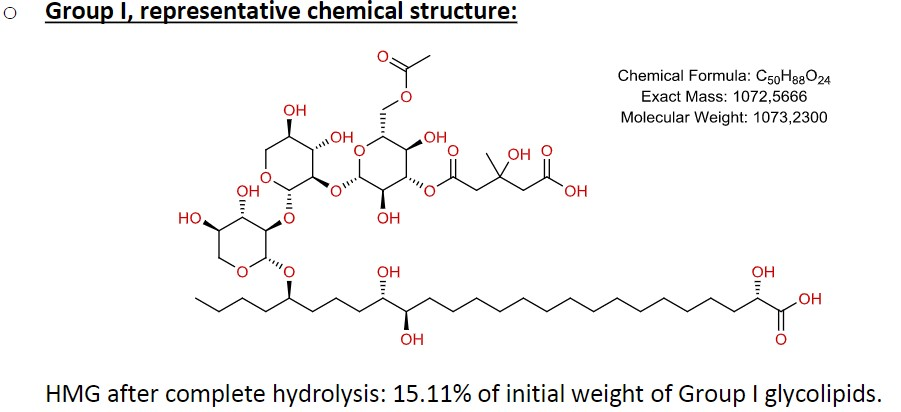
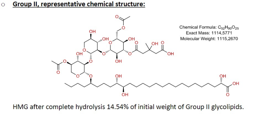
The maximum HMG amounts in percentage weight are calculated assuming a worst-case scenario of 100% hydrolysis of the acyl units, and assuming the maximum amount of HMG bearing glycolipids being present as fixed in the internal specification. Furthermore, this is based on a hypothetical glycolipid content of 100% in the product with water, protein, fat and sodium chloride content not considered, despite their actual presence lowering the total HMG content.
Internal specification of AM-1: <17% (sum of HMG-containing glycolipids Group I & II). Calculated theorical maximum amount of HMG obtained from AM-1: <2.52% (w/w) .
Based on HPLC-MS analysis data of the glycolipid composition batches used for the rat and ADME studies, dog and reproductivity toxicity studies, and a representative industrial scale batch were shown to have maximum HMG levels of 0.9% (w/w), 1.5% (w/w) and 1.7% (w/w) respectively after complete hydrolysis.
2.12.2. Chemical structure and identifiers of HMG
IUPAC Name: 3-Hydroxy-3-methylpentanedioic acid
Chemical Name: 3-Hydroxy-3-methylglutaric acid (HMG)
Synonyms: Dicrotalic acid, Mevalon, Medroglutaric acid
International Nonproprietary Name (INN): Meglutol
Chemical Abstracts Service Registry Number (CAS RN): 503-49-1
Registry of Toxic Effects of Chemical Substances (RTECS): MA3753000
The chemical structure of HMG is shown in Figure 16.
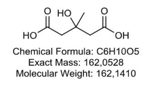
The Applicant claimed HMG is expected to be completely metabolised in the fatty acid pathway and tricarboxylic acid cycle like other dicarboxylic acids. HMG is a component of 3-hydroxy-3-methylglutaryl-coenzyme A (HMG-CoA) which is an intermediate in primary metabolism particularly in the mevalonate and ketogenesis pathways as well as leucine biosynthesis.
HMG is considered a naturally occurring constituent of human physiology and according to guidance in the FDA Redbook (1993) Concern Level paradigm, it would be classified as structure category A – of lowest concern.
The distribution of radioactivity after oral administration of 14C-HMG in male Swiss albino mice showed 14C-HMG was promptly and almost complete absorbed (Savoie & Lupien, 1975a). Radioactivity was observed in organs responsible for cholesterol synthesis (liver, intestinal wall) and with rapid and pronounced uptake of radioactivity also seen in the kidney. The authors concluded that the kidney is not only the main excretory route of HMG but also represents an important site of distribution for the compound (Savoie & Lupien, 1975a).
Toxicological investigations of HMG have been performed in pregnant rats and mice, (Savoie & Lupien, 1975b). Pregnant female Long Evans rats were administered daily intraperitoneal (i.p.) or oral gavage doses of HMG at 75, 250, and 500 mg/kg bw per day on Gestation Days (GD) 7-11. Pregnant female Swiss mice were administered Intraperitoneal injections (1600 mg/kg bw per day) or oral gavage (3700 mg/kg bw per day) doses of HMG on GD 8 and 10. Euthanasia and laparotomy were performed on approximately half of the pregnant rats on GD 21 while the remaining rats were allowed to deliver their offspring. All mice were terminated on GD 20 for performance of laparotomy. There were no effects of HMG treatment on reproductive performance of the dams, and no gross malformations of the fetuses or offspring were observed. The numbers of resorptions, percentage of live offspring after one month (evaluated in rats only), and fetal weights (reported for mice only) were not significantly different than controls (Savoie & Lupien, 1975b).
HMG was evaluated in 36 patients for its effect on plasma lipids in a double-blind human clinical trial (Lupien et al., 1979). Treatment groups were placebo, 0.750 g, 1.5 g, 2.25 g, and 3 g/day, there were no clinical or biological adverse effects due to the administration of HMG for 8 weeks. Hematologic, hepatic, renal, and metabolic function tests were clinically unaltered (Lupien et al., 1979).
Since HMG is an analogue of glutaric acid, glutaric acid provides additional information supporting the safety evaluation of HMG. When teratology studies were performed with glutaric acid in rats at 125, 400 and 1300 mg/kg and in rabbits at 40, 160 and 500 mg/kg, no adverse effects on pregnancy and no embryotoxic or teratogenic effects were observed (Bradford et al., 1984). Glutaric acid was tested in vitro in the Umu genotoxicity assay (Sakagami, et al., 1989) and in a rat in vivo bone marrow chromosomal aberrations assay (San Sebastian, 1989) at oral gavage doses as high as 2,750 mg/kg with negative results in both instances.
No adverse test-article related effects were observed in the toxicology and ADME studies (Bitzer, Henkel, Nikiforov, Rihner, & Henderson, 2017; Bitzer, Henkel, Nikiforov, Rihner, & Leuschner, 2017; Bitzer, Henkel, Nikiforov, Rihner, & Thomas, 2017; Herberth, 2017b, 2017a) performed using AM-1 test articles containing HMG at theoretical levels of 0.88% and 1.50% (calculated maximum theoretical amounts after complete hydrolysis based on measured amounts of HMG-containing glycolipids).
2.12.3. Potential Consumer Exposure to HMG
Assuming the unlikely event of complete hydrolysis the mean concentration of HMG can be calculated as 1.5% (w/w) as previously discussed, as glycolipids constitute approximately 93% of the AM-1 product and based on the exposure assessment previously conducted for AM-1, the potential dietary intake of HMG through consumption of formulated beverages containing AM-1 is calculated as follows:
For toddlers for which the highest exposure to AM-1 was identified (max. 95th percentile):
2.4 mg AM-1/kg bw per day x 93% glycolipids x 1.5 % (w/w) HMG = 33.5 μg HMG/kg bw per day.
For adults for the min. 95th percentile
0.547 mg AM-1/kg bw per day x 93% glycolipids x 2.52% HMG (w/w) = 7.6 μg HMG/kg bw per day.
However, excretion balance data from the rat ADME study indicates that AM-1 [14C] equivalents in urine and exhaled CO2 are less than approximately 20% of the administered dose (following an oral dose approximately 16-20% of recovered radioactivity was captured as expired CO2 and approximately 1% was in urine) and therefore, potential exposure to HMG is expected to be significantly lower, the exposure has been recalculated below:
For toddlers for which the highest exposure to AM-1 was identified (max. 95th percentile):
2.4 mg AM-1/kg bw per day x 93% glycolipids x 2.52% (w/w) HMG x 20% administered dose = 11.2 μg HMG/kg bw per day.
For adults for the min. 95th percentile
0.547 mg AM-1/kg bw per day x 93% glycolipids x 0.96% (w/w) HMG x 20% administered dose = 1.5 μg HMG/kg bw per day.
Both exposures are below the Threshold of Toxicological Concern (TTC) of 30 μg/kg bw per day which can be applied in the absence of genotoxicity and for HMG as a substance falling into Cramer Class I (according to EFSA and WHO Review of the Threshold of Toxicological Concern (TTC) approach and development of new TTC decision tree, published 10 March 2016).
The AEJEG recalculated exposures using the recalculated exposure values of 3.08 mg/kg bw per day for the toddler, which produced a highest exposure for toddlers of 72.2 µg/kg bw per day, which exceeds the TTC of 30 µg/kg bw per day for a Cramer Class I compound.
For toddlers for which the highest exposure to AM-1 was identified (max. 95th percentile):
3.08 mg AM-1/kg bw per day x 93% glycolipids x 2.52% (w/w) HMG x 20% administered dose = 72.2 μg HMG/kg bw per day.
The AEJEG also noted that the Applicant assumed 20% internal exposure (based on urinary excretion and CO2 exhalation), which is not typically accounted for in exposure calculations for TTC and would lower the exposure to 14.4 µg/kg bw per day.
If this approach was accepted, intakes for HMG would be below the TTC threshold. Nevertheless, the AEJEG noted that there were some uncertainties in adjusting for internal exposure in the use of TTC and that work in this area is ongoing. Therefore, whilst the concept of systemic dose/internal exposure was recognised as a relevant concept, together with a conservative assumption of 20 % internal exposure compared to what was reported in the study, it was recognised that there were still uncertainties in this approach.
2.12.4. Conclusion on HMG
The Applicant concluded HMG may be present as an acyl group of a minor portion of AM-1 glycolipids, meaning HMG may be theoretically freed via hydrolysis with an average theoretical concentration of <1.5% (w/w) in AM-1 batches. Results of organ distribution, reproduction, and developmental toxicity studies in two species (rat and dog) and a human clinical study support the safety of HMG. Furthermore, supporting teratology and genetic toxicity data on the structural analogue glutaric acid further supports the safety of HMG. Dietary intake calculations using conservative assumptions in potential exposures to HMG from use in AM-1 in the requested categories show these falls below the Threshold of Toxicological Concern (TTC). Considering all these factors, the presence of HMG as a potential hydrolysis product of a minor component of AM-1 is not a safety concern.
The AEJEG disagreed with the Applicant’s approach to use a 20% administered dose when calculating exposure to HMG for adults in the 95th percentile. An exceedance in the Threshold of Toxicological Concern (TTC) was also noted for toddlers using the updated exposure values.
2.12.5. Overall conclusion of the safety of the proposed use
The Applicant stated the in vitro genetic toxicity studies with AM-1 were all negative, confirming that AM-1, a glycolipid structure with no structural alerts for genotoxic metabolites has low potential for mutagenicity or carcinogenicity. AM-1 is poorly absorbed following oral intake and any that is absorbed appears to be rapidly hydrolysed to non-toxic compounds that are easily excreted. AM-1 hydrolysis products; glucose, xylose, acetate, isovalerate and long chain fatty acids are naturally occurring in the human diet and if absorbed may be ultimately metabolised to CO2.
Subchronic toxicity and multi-generation reproductive toxicity studies established high NOAELs with only minor non-adverse effects observed. These were concluded to be related to the surfactant and unpalatable properties of AM-1 and/or an artefact of the oral gavage route of administration.
Due to the well-defined ADME and PK profile of AM-1 in rodents, alongside the results of oral subchronic studies in both rats and dogs, it can also be concluded that chronic toxicity and carcinogenicity are not endpoints of safety concern for AM-1.
AM-1 and its ultimate hydrolysis product LCFA are poorly absorbed by the oral route and are primarily eliminated in the faeces without absorption. The Applicant stated absorbed components appear to be completely metabolized to CO2 and expired. There were no metabolites of safety concern identified for AM-1 or LCFA and no accumulation of these in tissues. Furthermore, the Applicant claimed, due to the rapid hydrolysis and elimination of AM-1 that occurs in vivo, the antimicrobial properties of the parent material would be absent or greatly diminished in the lower intestines so there is low concern for disruption of healthy gut microflora.
An in vivo rat study was submitted to investigate potential effects on the gut microbiota. Although assessment of microbiome-related outcomes remains an evolving area of science, the study did not identify substantial perturbations of the gut microbial ecosystem, including at exposure levels considerably higher than anticipated human dietary exposure.
AM-1 has low potential for systemic toxicity with oral repeated dose (90-day) NOAELs of ≥1200 mg/kg bw per day in rats (oral drinking water administration) and ≥ 1000 mg/kg bw per day in dogs (oral capsule administration), the highest dose levels tested, demonstrating that chronic toxicity and carcinogenicity are not of safety concern for AM-1.
AM-1 was determined to be non-genotoxic based on the results of an adequate range of in vitro genetic toxicity assays both in bacteria and mammalian cells including human lymphocytes.
AM-1 is not a reproductive or developmental toxicant as confirmed in the developmental and two generation reproduction study in rodents via oral gavage administration. Furthermore, the NOAELs of ≥1000 mg/kg bw per day determined by these toxicity studies are consistent with the NOAELs of the 90-day oral toxicity studies, providing additional confidence that there are no toxicological concerns.
2.13. Acceptable Daily Intake (ADI) for AM-1
The Applicant stated to be conservative the lowest NOAEL (1000 mg/kg bw per day) determined in the 90-day rat and dog toxicity studies would be utilised to derive of an Acceptable Daily Intake (ADI) for AM-1.
The Applicant used a default safety factor of 100-fold was used, based on an interspecies factor of 10 and an intra-species factor of 10. Due to the lack of chemical structural alerts, low oral bioavailability, simple and common hydrolysis products, and rapid and complete elimination of any metabolites; the Applicant did not consider an additional safety factor for the calculation of the ADI. The Applicant based their ADI on the 90-day toxicity study in Beagle dogs.
The AEJEG agreed with the use of the NOAEL of 1000 mg/kg bw per day, however, Members agreed on this level supported by the results of the 90 Day Rat study due to the study design and concluded this is more comparable with how humans would consume the product.
Members disagreed with the Applicant’s approach to the ADI and instead agreed an additional uncertainty factor of 3 to allow for extrapolation from a subchronic study to chronic exposure should be utilized as well as a factor of 10 for intra-species variation and 10 for inter-species variation. This gave a total uncertainty factor of 300, producing an ADI of 3.3 mg/kg bw per day, (Table 33).
The provisional ADI (3.3 mg/kg bw per day) established by the AEJEG was based solely on the toxicological data provided. The AEJEG did not consider the potential antimicrobial effects of the AM-1 and this aspect was not considered in establishing this ADI.
2.14. Documentation gathered
The use of glycolipids from Dacryopinax spathularia as a food ingredient was a new Application. The Applicant stated when performing literature searches for Dacryopinax spathularia in PubMed only 9 results were obtained, the 5 of these were publications by Bitzer et al. and were the only ones considered relevant for the safety assessment of AM-1 as a food ingredient (Bitzer, Henkel, Nikiforov, Rihner, & Henderson, 2017; Bitzer, Henkel, Nikiforov, Rihner, & Leuschner, 2017; Bitzer, Henkel, Nikiforov, Rihner, & Thomas, 2017). The AEJEG noted the Bitzer papers are written based on the study reports and testing of AM-1.
2.15. Safety evaluation strategy and corresponding testing strategy
The safety evaluation strategy and the corresponding testing strategy was built on EFSA’s Scientific Opinion Guidance for submission for food additive evaluations (EFSA, 2012). It included details on the chemistry and specifications of AM-1, lists and detailed existing authorisations and evaluations (US GRAS), and specified the proposed use of AM-1 as a preservative.
The toxicological studies for AM-1 include Tier 1- and Tier 2-testing as directed in the EFSA additive guidance (EFSA, 2012) (ADME, genotoxicity, acute toxicity, irritation and sensitization studies, sub-chronic toxicity, reproductive and developmental toxicity). There were no triggers for justifying the need for Tier 3-testing.
The safety assessment also included the edible jelly fungus Dacryopinax spathularia that is used to produce AM-1, this has a history of use as food, including being listed in the corresponding FAO compendium.
The exposure assessment calculated by the Applicant using EFSA’s Food Additive Intake Model (FAIM), both with normal use level, requested maximum use levels, individual use in the requested food categories and combined use in all food categories. Exposure from natural sources, food fortification, supplements or any other uses can be excluded as the there are no other known natural sources of the AM-1 glycolipids other than the producer organism (excluding its fruiting bodies).
2.15.1. Relevant published papers and unpublished studies including the individual raw data
The Applicant stated there were no new risk assessment data available which would affect the results of the existing risk assessments carried out.
The ADME and PK profile established for AM-1 in rodents, as well as the results of in vitro genetic toxicity studies with AM-1 and oral subchronic toxicity studies with AM-1 in rats and dogs, confirmed that chronic toxicity and carcinogenicity are not endpoints of safety concern for AM-1; therefore, chronic toxicity and carcinogenicity studies with AM-1 were not performed.
The Applicant claimed that available study results showed low oral absorption, no accumulation, and no toxicological endpoints of safety concern further support the conclusion that AM-1 exposure presents no health hazards to humans; therefore, a clinical safety study of AM-1 was not considered necessary for safety evaluation of the proposed uses. The Applicant claimed that as glycolipids such as AM-1 are part of the normal human diet and the fruiting bodies of the AM-1 production organism Dacryopinax spathularia had a history of use as food.
3. Conclusion
The FSA and FSS have undertaken a review of the AM-1 product using the scientific dossier provided, in addition to data relied upon by the applicant. The FSA and FSS concluded there were no toxicity, genotoxicity or other safety concerns for glycolipids produced using specifically the strain MUCL 53181 and agreed on a No Observed Adverse Effect Level (NOAEL) of 1000 mg/kg bw per day based on the 90-day rat study, and the use of a total uncertainty factor of 300 to give an Acceptable Daily Intake (ADI) of 3.3 mg/kg bw per day.
The FSA and FSS consider that there are no concerns over the safety of AM-1 for the proposed uses.
Abbreviations
Acknowledgements
The FSA and FSS would like to acknowledge Dr Allain Bueno, Prof. Qasim Chaudhry, Dr Olwenn Martin, Dr Claude Lambré, Dr Martin Rose, Dr Claire Stephenson, Dr Andrew Collins, Dr Carol Beevers and Prof. Shirley Price who worked on this application on the AEJEG.
The FSA would also like to acknowledge Members of the ACMSF and COT.