


Executive Summary
The advancement of rapid and accurate on-site diagnostic methods could provide tools to support risk management and decision making in the prevention and control of foodborne pathogens. Early detection is vital as it not only reduces threats to public health but also helps in mitigating foodborne illness outbreaks. The aim of this project was to identify promising technologies which could be piloted for on-site testing of foodborne pathogens. A comprehensive literature review was done to identify technologies under development for on-site testing. To ensure all relevant technologies were considered, the literature search was as broad as possible, encompassing both technologies proven or presumed to be applicable in detecting targets relevant to food safety, as well as those developed for other sectors. These technologies were assessed through a customised technology readiness level (TRL) framework with each technology assigned a TRL in combination with each in-scope pathogen or matrix. This led to the creation of a database of technologies and their respective readiness levels (TRL).
To understand the testing requirements in real world scenarios, an end-user study was conducted by creating a stakeholder map. This involved identifying both “strategic” and “operational” stakeholders, as well as organising focus groups and interviews. This study provided a broader perspective on on-site testing and helped in identifying sectors or processes that are well-suited or not suited for portable detection technologies.
Results from the literature review and end-user study led to the selection of four technologies shortlisted for their relative maturity and suitability for on-site use: portable real-time polymerase chain reaction (PCR), loop-mediated isothermal amplification (LAMP), lateral flow devices (LFD) and chemiluminescence kits. Data from end-user studies was used to identify suitable applications within the food sector for deployment of the technologies. Finally, a decision matrix allowed an evaluation of each of the shortlisted technologies in the context of specific end-user needs, and a final selection was made based on the outcome.
The two technologies taken forward for pilot on-site testing were portable real-time PCR for monitoring E. coli in irrigation water and LAMP for detection of Salmonella in high-risk foods not of animal origin at ports. Both technologies were at TRL 5 for in-scope pathogens and matrices. The testing methodology was initially validated in the laboratory, where a suitable real-time PCR assay for detecting E. coli and a LAMP test for Salmonella were identified. Following this, a DNA extraction method was developed, and various performance criteria such as specificity, sensitivity, repeatability, and reproducibility were evaluated. The finalised method was tested in the hands of end-users. Initially, training was provided to end-users at Fera, and subsequently, they conducted the tests themselves and provided feedback. Both methods showed good specificity with no observed cross-reactions for the selected assays. The sensitivity of the methods was not high enough to meet the strictest legislative requirements. Theoretically, the incorporation of enrichment steps could improve the sensitivity, though at the expense of making the process time-consuming. Also, a requirement from end-users was a simple, rapid test and no on-site handling of live cultures.
The end-users agreed that the portable real-time PCR for monitoring of E. coli in irrigation water was simple to use and overall feedback on usability of the method was positive. The accuracy of the test and certification based on the test results were considered as crucial factors for the implementation of this technology. Given the initial instrument cost of portable real-time PCR, it could potentially be offered as a service by agronomists, enabling the detection of various foodborne or plant pathogens. End-users from port health authorities agreed that implementing technologies like LAMP for on-site testing at ports requires infrastructure changes and additional staffing. They were able to conduct testing independently after the training. Factors such as test accuracy, diagnostic performance, and time to result were considered crucial for the adoption of the technology. To achieve full deployment of any technology, it is recommended to not only validate the method but also engage with end-users to gather feedback and tailor it to their requirements. This study may also suggest requirements for changes in legislation and infrastructure to support the implementation of these technologies.
Overall, the study currently supports a role for rapid on-site diagnostics as complementary tools within food systems, where their strengths in speed and accessibility can enhance risk management and decision-making, provided their limitations are clearly understood and communicated.
1. Introduction
According to the Alert and Cooperation Network 2022 Annual Report (Commission, 2023), pathogenic microorganisms were the second most reported hazard category in food (857 notifications) after pesticide residues (990 notifications). The most reported pathogenic microorganism was Salmonella, followed by Listeria monocytogenes and Escherichia coli. These pathogens are among the Food Safety Agency (FSA) priorities, and part of the scope of the Pathogen Surveillance in Agriculture, Food and Environment (PATH-SAFE) Programme. Detecting foodborne pathogens could help safeguard public health, ensure consumer safety, protect economic interests, and maintain the reputation of food businesses. Prompt detection plays an important role in monitoring, verification, and response to minimise the risks associated with foodborne illnesses.
Point-of-care (POC), on-site or in-field diagnostics for foodborne pathogens could play an important role in safeguarding public health and ensuring food safety. These rapid and efficient testing methods could be indispensable in identifying harmful pathogens like Salmonella, E. coli and Listeria at the earliest stages of food production and distribution. By providing quick and on-site results, they will enable interventions to support risk management and reduce the risk of foodborne illnesses and outbreaks. On-site diagnostics will allow food manufacturers, distributors, inspectors and regulators to make informed decisions, implement appropriate control measures, and maintain the integrity of the food supply chain. Recently, effort has been made to develop rapid diagnostic technologies for direct on-site testing, which offer greater adaptability and ease of use over conventional methods which require well-trained personnel in laboratories, bulky equipment and extended processing times. However, for the adoption of on-site diagnostic methods, they need to follow the ASSURED criteria (Kosack et al., 2017):
-
Affordable, cost-effective to ensure large business and small producers can adopt these methods.
-
Sensitive, able to detect low levels of pathogen to ensure food safety.
-
Specific, able to differentiate e.g., pathogenic vs non-pathogenic strains.
-
Rapid, providing quick results to allow immediate action if required.
-
Validated, to allow results to be properly interpreted.
-
Portable, to allow easy transportation and storage.
-
Integrated, providing sample-to-answer processes, and streamlined information sharing with e.g., authorities.
-
Accepted by regulations, comply with standards, and be accepted by relevant authorities.
This project formed part of PATH-SAFE Work Stream 3a (WS3a). As part of this project, a literature review study (Work Package 1) to identify promising technologies with on-site testing applications was performed (Figure 1). To gather all possible relevant technologies, the literature review had to be as broad as possible to include both technologies with demonstrated or presumed applicability to detection/diagnosis of targets of relevance to food safety and those which have been developed for use in other sectors, such as clinical and veterinary science, agriculture, plant health or environmental monitoring. These technologies were assessed through a customised technology readiness level (TRL) framework, to determine the maturity of the technology and its readiness for deployment for in-scope pathogens and matrices. This new TRL framework tackles some of the ambiguities that previous published TRL frameworks showed, as most remain heavily based on the generic level descriptions originally developed by NASA. For example, the use of terms ‘validation’ and ‘demonstration’ in the definitions of TRLs. While interpretation of these terms can vary, ‘validation’ of functionality in the context of this project involves confirming the ability to detect a target pathogen at a specified level, confirming that non-target pathogens are not detected, and determining the frequency with which false positive and/or negative results can be expected to occur. ‘Demonstration in a relevant/operational environment’ involves confirming that the technology under consideration can be combined successfully with any other components necessary for in-field testing, and that the same performance can be achieved under field conditions as well as in the laboratory. A TRL was assigned to individual technologies in combination with each in-scope pathogen or matrix, as the technologies had reached different levels of development for each target. The database of technologies with TRL assignments supplied a snapshot of the maturity of each technology, and an indication of the next steps to progress towards deployment in a particular application.
As part of the project, an end-user study was performed (Work Package 2). Several focus groups and interviews with strategic stakeholders and operational end-users were conducted. The main aim of these interviews was to provide higher level context for on-site testing and gain insights into any sectors or processes which may be either particularly suitable or unsuitable for the use of portable detection technologies. On-site diagnostic methods are designed to provide rapid solutions to address real-world challenges. Thus, end-user feedback is key to understand if technologies meet the specific needs and the preferences of those who rely on them daily.
Outcomes from the first two work packages led to a pilot study (Work Package 3), aiming to trial on-site testing of two rapid diagnostic technologies with end-users (Figure 1). A shortlist of the technologies with the most potential to take forward for a pilot study with end-users was decided. Many promising technologies were discounted due to the low chance of progressing past early TRLs. Validation of the chosen technologies in the laboratory was performed, as well as training with the end-users and independent testing.

2. Materials and Methods
2.1. Scope
The scope of the literature review, and the subsequent technology readiness level (TRL) assessment, was defined in terms of the target pathogens of interest, the sample types which may be tested, and the range of settings in which testing could be carried out.
2.1.1. Pathogens of interest
The literature review considered technologies with the potential to be applied to on-site testing scenarios of relevance to PATH-SAFE, regardless of the targets for which they were originally developed or for which they have been applied to date. The following list of pathogens of interest (Table 1), however, allowed consideration of specific test characteristics that were required in the assessment of the suitability of technologies for the applications under consideration.
2.1.2. Sample types
A broad range of matrices were within the scope of the landscaping study, including (but not limited to) to those described in Table 2.
2.1.3. Settings for testing
Definitions for on-site testing can encompass settings ranging from ‘field’ situations with no specific facilities where the conditions may be relatively challenging to rudimentary ‘laboratory’ facilities, for example, as could be envisaged at ports of entry. Potential skill levels of operators will also vary, together with the time and resources available to them. Technologies appropriate for all settings (and operator skill levels) along this continuum were in scope for the literature review.
2.1.4. Technologies
All technologies with the potential to be applied to detection of the pathogens of interest listed above (2.1.1) were within scope for the literature review. This will include both complete testing solutions (sample to result), where these exist, and components which could be combined to produce suitable testing solutions (e.g. extraction methods, detection methods, instruments/engineering solutions).
2.2. Literature review
An extensive literature search was undertaken to identify technologies which are being developed for on-site testing. Searches were performed, using both Web of Science and Scopus as search engines, using the ‘All Databases’ option. Firstly, a search focusing on known on-site technologies applied for the detection of foodborne pathogens was performed using Web of Science. Subsequently, a second search focusing on novel on-site technologies, aiming to identify methods applied in different fields such as veterinary or clinical science, was performed using both search engines. A final search focusing on crude extractions was performed using Web of Science. Prior to performing the searchers, key search terms were identified and defined, as well as the right combination of Boolean operators. Publication year was limited to a range between 2018-2023, to identify the most recently published articles. The final search terms employed for all searches were as follows:
-
Direct search of in-field foodborne pathogen detection methods:
Topic:
(“Food” OR “Foodborne” OR “Food-borne” OR “Food borne”) AND
(“on site” OR “in-field” OR “in field” OR “point of care” OR “point-of-care” OR “in situ” OR “point-of-need” OR “on-site” OR “pen-side” OR “onsite”) AND
((“diagnos*” OR “detect*” OR “identif*”) AND (“method*” OR “test*”)) NOT
(“allergen*” OR “mycotoxin*”)
Year published:
-
Language:
English
-
-
Search of novel in-field detection methods applied in different disciplines:
Topic:
(“on site” OR “in-field” OR “in field” OR “point of care” OR “point-of-care” OR “in situ” OR “point-of-need” OR “on-site” OR “pen-side” OR “onsite”) AND
((“diagnos*” OR “detect*” OR “identif*”) AND (“method*” OR “test*”)) AND
(“novel” OR “emerg*” OR “new*”) AND
(“Bacteri*” OR “Viru*” OR “Fung*” OR “DNA” OR “RNA” OR “Nucleic acid”)
Year published:
2018-2023
Language:
English
-
Search of crude extraction methods
Topic
(“Nucleic acid” OR “DNA” OR “RNA” OR “sample prep*”) AND (“on site” OR “in-field” OR “in field” OR “point of care” OR “point-of-care” OR “in situ” OR “point-of-need” OR “on-site” OR “pen-side” OR “onsite” OR “equipment free”) AND (“Water” OR “Meat” OR “Shellfish” OR “Dairy” OR “Swabs” OR “Animal feed” OR “Fish” OR “Fresh Produce” OR “RTE” OR “Ready to eat”) AND (“Bacteria” OR “Virus”) AND (“extract*”)
Year published:
2018-2023
Language:
English
Once literature searches were performed, results were downloaded into EndNote and RIS files, and the latter were imported into Covidence (https://www.covidence.org), a literature review management software. Duplicated references were automatically discarded by the software after the import step. Within Covidence, curation of the references was performed in two main steps: a title and abstract screening to quickly discard those publications that were irrelevant, and a second step to look at full text of relevant publications and obtain the relevant information for the TRL assessment of each technology. During the title and abstract screening step, publications were labelled using study tags. Those study tags were both general such as ‘DNA-based detection’ or ‘phage-based detection’ and technology-specific such as ‘PCR’ or ‘NASBA/Nucleic Acid Sequence Based Amplification’. The use of study tags allowed grouping of publications by technology in the full text review stage to obtain the relevant information on each technology for the TRL assessment. A record of results on each step of the search was maintained.
Due to the large volume of publications within this second phase of the literature review, review papers for each technology were prioritised, as they could potentially gather most of the information available or indicate relevant papers to read for each of the technologies. Subsequently, the prioritised publications to read were those describing work on foodborne pathogens or pathogens in-scope. The next step was to review those papers where the technology had been applied to pathogens outside scope, but where the technology was taken forward and performed e.g., on-site testing, or testing with relevant end-users, which would make the technology progress in the TRL framework. All the relevant information for evaluation of the technologies was gathered, and each technology was assigned a TRL.
In addition, further sources were also explored. A search was performed to identify any other relevant commercial kit available by looking into company webpages such as Creative Diagnostics, Romer Labs or Merck Millipore. In addition, the USDA has published a list of foodborne pathogen test kits validated by independent organisations (Food Safety and Inspection Service, U. S. D. o. A., 2020), including kits for Salmonella, Listeria, Escherichia coli and Campylobacter, which were assessed.
2.3. Technology Readiness Level (TRL) framework
A TRL framework was developed as part of the project (Figure 2 and Table 3). This TRL framework was used to assess the individual technologies listed in the database after performing the literature review in combination with each in-scope pathogen or matrix (Annex 1). TRL 1 and 2 were beyond the scope of the literature review as technologies at these early levels of development were unlikely to be captured by the study’s search terms. The database of technologies with TRL assignments supplied a snapshot of the maturity of each technology, and an indication of the next steps to progress towards deployment in a particular application. To perform the assessment, a TRL tool was developed, including several questions in each TRL level that needed to be fulfilled to reach each respective level (Appendix A).
_framework_developed_as_part_of_path-safe_ws3a.svg)
2.4. End-user study
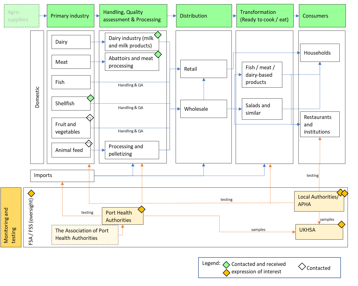
To inform the end-user study, the project used the target pathogens and sample matrices defined at the outset to determine key areas where pathogen can be expected along relevant supply chains (Table 4, 5). These were used to create a base map of key stakeholder groups. In a last step, the map was expanded to stakeholders from the regulatory environment (Table 6; Figure 3).
A contact database was created using existing connections of the Food Standards Agency (FSA) and Fera Science Ltd. Where necessary, the database was expanded using a snow-balling approach. To enable focused stakeholder engagement, the database was split into ‘strategic’ and ‘operational’ stakeholders. Nine strategic stakeholders from FSA, APHA, DEFRA, CEFAS and UKHSA were consulted first, to provider wider context and information on past efforts. To delve into specific end-user needs, six operational stakeholders including representatives from port health authorities, retailers, local authorities and food businesses were consulted. The initial plan was to conduct workshops; however, this had to be adjusted for logistic reasons. In the end, the team conducted several focus groups and interviews. In addition, presentations to a project network and the steering group were used for additional feedback. Additional interviews were held on the back of both meetings.
Focus groups and interviews aimed to provide higher level context for on-site testing, as well as gain insights into sectors or processes where portable detection technologies might be most or least applicable. Based on these insights, the team identified key scenarios and used them to develop a selection matrix bringing together end-user feedback, technology insights using results from the literature review, and policy feedback on potential future impacts. The selection of case studies was largely influenced by the sectors where we were able to secure engagement with.
2.4.1. Map of key stakeholders
A stakeholder map (Figure 3) was built around the scope of target pathogens and samples determined at the beginning of the project, briefly summarised in Table 4 and Table 5. More specifically, an initial assessment of the main impact points of pathogens, in combination with key supply chains, was used to create a map of key stakeholder groups. This was then expanded to include stakeholders from the regulatory environment (Table 6).
2.5. Pilot study
The two technologies to be taken forward for the pilot study (Work Package 3) were:
-
Portable real-time PCR for monitoring of E. coli in irrigation water.
-
LAMP for detection of Salmonella in sesame seeds at ports.
In phase one of Work Package 3, we contacted appropriate stakeholders from Work Package 2 to select the testing location and secure their engagement in the project. The aim was to organise visits to testing locations and consult end-users to assess facilities available for testing and inform test development. Both technologies selected were at TRL 5, as although assays were developed for the target pathogens, they had not been demonstrated with all the components needed for testing. For the detection of E. coli in irrigation water, the important steps were to select a i) suitable water filtration method, ii) DNA extraction method, and iii) real-time PCR test and evaluate these components together. Similarly, for detection of Salmonella in high-risk food not of animal origin (HRFNAO), a suitable i) sample preparation method, ii) DNA extraction, and iii) LAMP test were identified and test performance evaluated in the laboratory. Once methods were optimised, small scale testing in the laboratory was performed with a panel of spiked and real samples to confirm expected performance compared to a gold standard method.
In phase two of Work Package 3, technology transfer to end-users began. First, training was provided for end-users at Fera Science Ltd. Secondly, supervised on-site testing was carried out with end-users for the irrigation water scenario, and unsupervised on-site testing was performed by the Port Health Authorities. Samples were tested to determine end-users understanding and competency in the method. Visits and sessions with the end-users led to discussion to gain feedback on the test and technology. Where possible, samples tested on-site by end-users were tested in parallel with a gold standard method in the laboratory.
2.5.1. Detection of Escherichia coli in irrigation water
2.5.1.1. Real-time PCR assay selection
Three real-time PCR assays for the detection of E. coli were selected from the literature for comparison (Silkie et al., 2008; Srinivasan et al., 2011; Walker et al., 2017). The E. coli UidA assay from Silkie et al. (2008) was run as follows, reaction mixtures consisted of 12.5 μl of Environmental Master Mix, 600 nM of each primer and 300 nM of the probe, made up to 24 μl with molecular grade water, and 1 µl of template DNA. Cycling conditions were 95 °C for 10 minutes and 40 cycles of 95 °C for 10 seconds and 60 °C for 1 minute. The E. coli Srinivasan et al. (2011) assay reaction mixture consisted of 12.5 μl of Environmental Master Mix, 600 nM of each primer and 200 nM of the probe, made up to 24 μl with molecular grade water, and 1 µl of DNA template. Cycling conditions were 95 °C for 10 minutes and 40 cycles of 95 °C for 10 seconds and 60 °C for 1 minute. In the E. coli Walker et al. (2017) assay each reaction consisted of 12.5 µl of IQ SYBR Supermix reaction buffer, 300 nM of each primer, made up to 24 μl with molecular grade water, and 1 µl of template DNA. Cycling conditions were 95 °C for 2 minutes followed by 40 cycles of 95 °C for 15 seconds, 68°C for 60 seconds. Followed by a high-resolution melting curve analysis.
An initial comparison of the assays analytical sensitivity used E. coli pure culture (smsco004) grown on nutrient broth for 24 hours and then heat treated at 95°C for 5 minutes to lyse the cells. The heat-treated pure culture of E. coli was tested from neat to 10-6 dilution with two technical replicates per dilution tested on each real-time PCR assay. The most sensitive assay from the initial test was taken forward for comparison with the commercial kit BioPoo® E. coli RT-PCR Go-Strips® (Biomeme) for full assessment of analytical sensitivity and specificity.
DNA was extracted from two isolates of E. coli (522-036052, 522-036341) using the DNeasy Blood and Tissue Kit (Qiagen), following the manufacturer’s instructions. DNA concentration was quantified using the Qubit™ dsDNA Quantification Assay Kits (Invitrogen™). The DNA extracts were diluted to a starting concentration of 10 ng/µl, and then a 10-fold dilution series was created to 1 fg/µl. Each dilution series was tested on the commercial kit and the Silkie et al. assay with two technical replicates per dilution. When testing extracts using the commercial kit 19 µl of molecular grade water was used to resuspend the dried reagents, followed by 1 µl of sample DNA. The Silkie et al. assay sensitivity was additionally tested on the Franklin® thermocycler under standard run conditions, but with 25 µl of mineral oil above the reaction to prevent evaporation to determine any differences associated with real-time PCR instruments.
Isolates of target and non-target bacteria were acquired from Fera’s culture collection and University of Lincoln (Table 7). The cultures were grown on nutrient broth for 24 hours and then heat treated at 95°C for 5 minutes to lyse the cells and release DNA. The DNA extracts were 10-fold diluted and used directly to test the specificity of the real-time PCR assays. Additional DNA extracts from isolates of E. coli isolated from river water were kindly provided by Centre for Environment, Fisheries and Aquaculture Science (CEFAS) to improve the inclusivity panel and were only tested on the commercial kit BioPoo® E. coli RT-PCR Go-Strips® (Biomeme) on the Franklin® thermocycler (Biomeme). When testing extracts using the commercial kit 19 µl of molecular grade water was used to resuspend the dried reagents, followed by 1 µl of sample DNA.
2.5.1.2. Sample preparation and DNA extraction
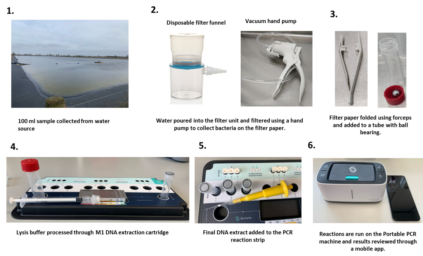
Sterile distilled water spiked with E. coli to a concentration of 104 CFU / 100 ml, was used to assess all methods. To create quantified samples of E. coli, an isolate (S22-035750) was grown on nutrient agar for 24 hours at 37°C, purity was assessed visually and then sub-cultured into nutrient broth for 24 hours at 37°C. The nutrient broth was quantified on a spectrophotometer at 280 nm and diluted to 0.1 optical density which was determined to be around 108 CFU/ml and a dilution series was made from here to 104 CFU/ml. Plate counts were used to confirm the concentrations. A 1 ml sample of the 104 concentration nutrient broth was used to spike a 100 ml sterile water sample. The Silkie et al. (2008) assay was used to test the DNA extracts from each method to compare Ct values.
Two methods of water filtration to concentrate the bacterial cells within the 100 ml sample volume were assessed. Filtration method A1 involved a single use analytical filter funnel (Nalgene™) with a 0.45 µm pore filter, used in combination with a hand-operated vacuum pump (Nalgene™) to filter the sample. The filter paper was removed, and DNA extracted using the DNeasy PowerWater kit (Qiagen) following the manufacturer’s instructions. In filtration method A2, the sample was filtered by adding it to the column of a 50 ml plastic syringe, with a syringe filter (0.45 μm pore size) (Nalgene™) attached. The water was processed in two 50 ml batches and passed through the filter to collect the bacterial cells. After the sample has been processed the syringe filter was moved onto a 10 ml syringe and washed with 10 ml of sterile water passed through the filter. Finally, the filter was moved onto a 1 ml syringe and the cells removed from the filter by backflushing with 200 µl of molecular grade water drawn through the filter into the syringe. The filter is then discarded, and DNA extracted from the 200 µl sample using the DNeasy PowerWater kit (Qiagen) following the manufacturer’s instructions.
Equipment free DNA extraction using the M1 Sample Prep Cartridge DNA-HI (Biomeme) was compared to the DNeasy PowerWater kit (Qiagen) as the gold standard. In short, after water filtration using method A1, described above, the filter paper was loosely rolled and added to a 5 ml tube with a stainless-steel ball bearing added to the centre of filter paper, 2 ml of BLB lysis buffer (Biomeme) was added and cells disrupted through manual shaking for one minute. Following this the sample was processed through the M1 Sample Prep Cartridge following the manufactures instructions.
2.5.1.3. Assessment of test performance
The final test for validation was as follows: Samples were filtered using single use analytical filter funnel (Nalgene™) with a 0.45 µm filter, used in combination with a hand-operated vacuum pump (Nalgene™). The filter paper was removed, loosely rolled, and added to a 5 ml tube with a stainless-steel ball bearing added to the centre of the filter paper. Cells were disrupted by adding 2 ml of BLB lysis buffer (Biomeme) and manual shaking for one minute. Following this, the sample was processed through the M1 Sample Prep Cartridge following the manufactures instructions, and 20 µl of the final DNA extract was added to the commercial kit BioPoo® E. coli RT-PCR Go-Strips® (Biomeme) and mixed by pipetting to resuspend the dried reagents. The PCR was run on the Franklin® Thermocycler (Biomeme) following the manufacturer’s instructions (Figure 4).
The analytical sensitivity of the final method was assessed using a dilution series of E. coli (S22-035750) grown in nutrient broth and quantified as described previously. The samples created ranged between 106 CFU/100 ml and 101 CFU/100 ml, using sterile distilled water. The repeatability of the method was assessed by processing three samples near the determined limit of detection of the method, the samples were then tested in triplicate real-time PCR reactions. A second user then repeated this to determine reproducibility.
To determine the diagnostic performance of the test on real samples irrigation water sources were identified by end users and samples collected and tested by real-time PCR method, and the gold standard method for enumeration of E. coli in water through membrane filtration and confirmation (ISO 9308-1:2014). The location of sampling was recorded using “what3words”, along with the type of water source and the crop types which the water source is used to irrigate (Table 8). Samples were sent by overnight courier to the lab and tested within 24 hours of collection. Results from parallel testing were used to calculate diagnostic sensitivity, diagnostic specificity, positive predictive value, and negative predictive value of the test. Anything with >100 CFU/ 100 ml as determined by the golden standard method was considered positive for potential contamination and anything below this considered negative.
Potential inhibition of the real-time PCR by the real samples was investigated through spiking some of the samples with E. coli, diluting samples and adding 1 µl (50 mg/ ml) bovine serum albumin (BSA) to the reactions.
2.5.1.4. Training with end-users
Two end-users were trained to use the technology as part of the project. The training was conducted in two stages. The first stage was performed at Fera where the end-users were provided with a protocol describing the method and how to perform it (Annex 2). The method was then demonstrated to the end-users, before they undertook the procedure themselves with negative water samples under supervision. The second stage involved meeting the end-users at an on-site location to perform the testing on real irrigation water samples in a non-laboratory environment under supervision. Positive and negative controls were included to account for potential contamination. As part of parallel testing, duplicate water samples and extracted DNA tested on-site were also analysed in the laboratory to compare the PCR results. Results from on-site testing were also compared to those obtained using the gold standard method. A feedback questionnaire was sent to end-users to get their perspective on the technology (Annex 3).
2.5.2. Detection of Salmonella in sesame seeds
2.5.2.1. LAMP assay selection
Three LAMP assays were selected for Salmonella testing, assay 1 (Ge et al., 2019), assay 2 (D’Agostino et al., 2015) and assay 3 (BK-S. enterica-050, OptiGene Ltd.). For assay 1 and 2, the LAMP reaction mixture in a total volume of 25 µl contained 15 µl 1x isothermal master mix ISO-004, 0.2 µM F3 primer, 0.2 µM B3, 2 µM FIP, 2 µM BIP, 2 µM F-loop, 2 µM B-loop, 2.5 µl sterile nuclease-free water and 5 µl of DNA template. Assay 3 was obtained from OptiGene Ltd. in the form of a kit containing ISO-004 and primer mix. Due to proprietary rights, primer information for this assay was not available. For assay 3, the reaction contained 15 µl isothermal Mix 004, 5 µl primer mix and 5 µl DNA template. The LAMP reaction run parameters were amplification at 65°C for 20 minutes followed by annealing from 95-75°C with a ramp rate of 0.05°C/s.
Pure cultures of Salmonella were grown on nutrient broth at 37 °C for 24 hours. For initial comparison of the analytical sensitivity of the assays, a heat-treated (95°C for 5 minutes) pure culture of Salmonella Typhimurium smscoo1 was used from neat to 10-8. The assays showing the most promising results in the preliminary sensitivity experiments were further evaluated for analytical sensitivity and specificity.
To determine the analytical sensitivity of the LAMP assay, the DNA from two isolates, Salmonella Enteritidis 024793_5/5 and Salmonella sp. 023777_1/4 was extracted by heating treatment and diluted from neat down to 10-8 to create a 10-fold dilution series. Each dilution was tested with assay 1 and assay 3.
For evaluation of specificity, isolates of target and non-target bacteria were obtained from Fera’s culture collection and the University of Lincoln (Table 9). To lyse the bacterial cells, 1 ml of broth was heated treated and diluted down to 10-3 using nuclease free water and 5 µl volume was used in the LAMP reaction. Additional DNA extracts from isolates of Salmonella isolated from river water were kindly provided by the Centre for Environment, Fisheries and Aquaculture Science (CEFAS) to improve the inclusivity panel and were only tested on the assay 3.
2.5.2.3. Development and testing of crude extraction methods.
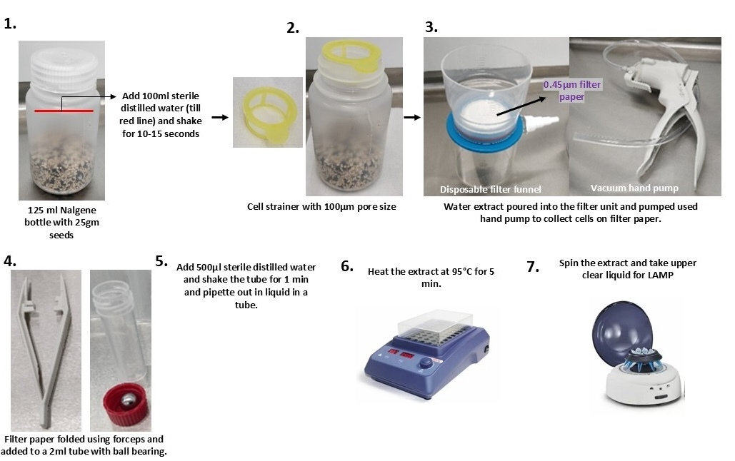
A total of 25 g of sesame seeds were spiked with 1 ml broth containing 103 CFU/ml of Salmonella sp. 023777_1/4 directly into a 125 ml Nalgene bottle. The culture method and quantification procedure were as described in section 2.5.1.1. A variety of sesame seeds such as roasted mixed, white, brown, and black were included in the experiments. Five replicates were tested for each method using assay 3.
To transfer bacterial cells from the surface of the sesame seeds into suspension, 100 ml of sterile water was added to the Nalgene bottle. The bottle was manually shaken by hand for 10 to 15 seconds to dislodge bacterial cells from the seed surface into the aqueous phase. In Method B1, no filtration or clarification step was performed. Instead, 1 ml of the resulting seed wash water was taken directly from the bottle and subjected to heat lysis at 95°C for 5 minutes. Following heat treatment, 5 μl of the lysate was added directly to the LAMP reaction mixture.
Next, filtration-based techniques were evaluated. To separate the seeds and water, a 100 µm pore size cell strainer (Scientific Laboratory Supplies) was fitted to the top of the Nalgene bottle and water was poured directly into an analytical filter funnel (Nalgene™) equipped with a 0.45 µm pore size filter paper. A hand-operated vacuum pump (Nalgene™) was used to filter the samples and concentrate bacterial cells onto the filter paper. The filter paper was then loosely rolled and inserted into a 5 ml tube with a stainless-steel ball bearing.
In method B2, 500 µL sterile water was added to the centre of the 5 ml tube and vigorously shaken for 1 minute to dislodge bacterial cells attached to the filter paper into the water. Subsequently, 250 µl of this extract was pipetted into a 1.5 ml tube and heated at 95°C for 5 minutes on a heating block. Following heating, samples were briefly spun to ensure any particulate matter settled at the bottom and the upper clear solution was then used in the LAMP reaction.
In method B3, a Bento dipstick DNA extraction kit was used after filtration. 500 µL extraction buffer was added to the 5 ml tube containing the filter paper and a stainless-steel ball bearing and shaken for 30 seconds. The dipstick was dipped three times in the extraction buffer, followed by 5 times in the wash buffer. Finally, DNA was released from the dipstick into the LAMP reaction strip containing reagents by dipping it 3-15 times.
The crude extraction methods were also compared with the standard laboratory DNA extraction method. In method B4, following filtration, the filter paper containing bacterial cells was processed using the Qiagen DNeasy Blood & Tissue Kit extraction following the manufacturer’s instructions.
2.5.2.3. Assessment of test performance
The final crude extraction method is represented in Figure 5.
The analytical sensitivity of the overall method was assessed using a dilution series of Salmonella sp. 023777_1/4 grown in nutrient broth and quantified as described in section 2.5.1.1. The dilutions ranging from 105 to 101 CFU/ml were made in sterile distilled water and 1 ml volume was added to 25 g of sesame seed samples. To estimate the loss of sensitivity during the DNA extraction process, 1 ml broth of same dilutions was heated at 95°C for 5 minutes, and 5 µl volume was tested in the LAMP reaction.
To evaluate the repeatability of the method, three 25 g seed samples were spiked with Salmonella sp. 023777_1/4 culture near limit of detection (LOD) and three technical replicates were tested in the LAMP reaction. To account for reproducibility, another user replicated the entire process in similar manner.
The performance of overall methodology was also accessed on real samples. Our team visited port health authorities during sampling sessions to gain insight into the operations and procedures at the port, including inspection protocols, regulations, and testing requirements. As part of the regulatory testing, Suffolk Coastal Port Health Authorities in Felixstowe test sesame seed shipments for Salmonella through the UK Health Security Agency (UKHSA). To evaluate the methodology developed in this pilot study, sub-samples from the samples submitted for actual testing were sent to Fera science ltd. via overnight courier. The sample number, country of origin and variety of sesame seeds were recorded. Testing results from UKHSA were compared with the results obtained from this study.

2.5.2.4. Training with end-users
Two port-health officials from Suffolk Coastal Port Health Authorities, Felixstowe were trained to use the technology. The training was conducted in two stages. The first stage was carried out at Fera where the end-users were provided with a detailed training instructions describing the method and how to perform it (Annex 4). The method was first demonstrated for end-user followed by end users performing the method themselves with negative seed samples.
Second stage involved on-site training at the port followed by end-users independently performing the test for one month. Positive and negative controls were included in the testing to account for potential contamination. As part of parallel testing, seed samples tested on-site were also tested in the lab. Furthermore, DNA extracted by end-users on-site was also analysed in the lab to compare the LAMP results. Results from on-site testing were compared with the results from the UKHSA as gold-standard testing. A feedback questionnaire was sent to end-user to get their perspective on the technology (Annex 5).
3. Results
3.1. Literature review of on-site diagnostic technologies
The literature review was conducted by performing a search focusing on known on-site technologies, a second search focusing on novel on-site technologies, and a search focusing on crude extractions. The results of these searches were as follows:
-
Direct search of on-site foodborne pathogen detection methods: 5,870 hits from a search performed in December 2022 in Web of Science.
-
Search of novel on-site detection methods applied in different disciplines: 13,196 hits from a search performed in December 2022 in Web of Science and 7,200 hits in Scopus in February 2023.
-
Search of crude extraction methods – 267 hits February 2023.
A total 19,920 results were screened for title and abstract using Covidence at the first step. Several publications were excluded for the following reasons:
-
Described a non-relevant target (toxins, chemicals etc.).
-
Technology is not possible to use in-field.
-
Did not described a detection method.
-
Full paper was not accessible.
From the remaining pool of publications, 2,785 were split by technology type using the study tags created and further evaluated at full text level in the second stage of the literature review. Information on each technology was gathered to perform the TRL assessment. These results allowed the creation of a large database of technologies as well as their TRL assessment (Annex 1).
After further evaluation of other sources outside publications, no further suitable kits for on-site testing were identified either on corporation webpages or the validated test list published by the Food Safety and Inspection Service (2020).
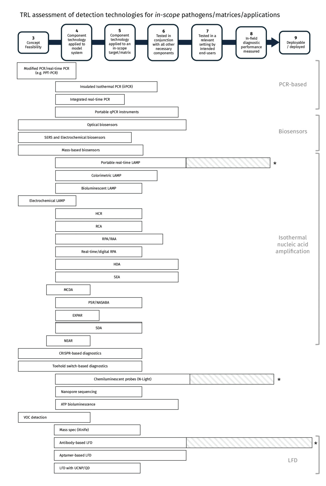
Below the results of the literature review are described, including a technical overview of each of the technologies assessed and the TRL assessment. Figure 6 shows a summary of TRL assessments for testing of in-scope pathogens, matrices and/or applications; although some technologies have been operationally deployed for out-of-scope targets, this is not represented in Figure 6. For all technologies, the TRL assessment is represented as a range of levels extending from TRL 4 (in some cases 3) because the technology has been applied to some of the in-scope pathogens/matrices but not others (e.g., a test has been reported for Salmonella but not for Listeria); no individual technology has been applied to all in-scope pathogens and sample types. The TRL assessment described in the literature review represents the most advanced examples demonstrated in reviewed publications.
It should be noted that in the context of this TRL framework, a commercially available diagnostic device or kit, or a method with evidence of operational use, would not necessarily be assessed as TRL 9, because application-specific validation data is required to determine whether a test can be usefully deployed in a particular scenario. Some commercially available technologies could have reached TRL 8-9 in application-specific scenarios, however, the publicly available evidence was missing to fully support the TRL. As may be noted from Figure 6, while basic data to verify certain aspects of test performance (often analytical sensitivity and specificity) are available for many tests, diagnostic performance data is generally lacking or is not applicable to relevant testing scenarios, for example, validation data has been collected in the laboratory rather than on-site conditions. Even for the most mature and established technologies, a necessary step in the progression towards deployment for a particular purpose is therefore the collection and analysis of application-specific validation data.
Commercial availability, however, may be a requirement for achieving TRL 7, as accessibility of instruments, devices and/or reagents is a prerequisite for meaningful testing by end-users to take place. Some assessment of technical performance under field conditions may be possible using, for example, a prototype device or custom prepared reagents, but assessment of the reliability of production-model devices and reagents, and consideration of real-world logistics (such as reliability of supply chain, storage requirements, etc) are also necessary.
Many of the technologies shown in Figure 6 are based on established concepts which have been applied to detection of one or more target organisms (TRL 4), some of which may be within the scope of this project (TRL 5). However, some of the more broadly defined technological areas remain the subject of activity at TRL 3, in which new concepts are being developed and tested for feasibility. Development of biosensors and test components based on synthetic biology (for example, CRISPR, toehold switches) are areas of on-going diagnostic innovation.
_reached_for_each_technology_for_testing_of.png)
3.1.1. Polymerase chain reaction (PCR)
Polymerase chain reaction (PCR) is the most established method for nucleic acid amplification and remains the gold standard for pathogen detection. Its strengths include high analytical sensitivity and specificity, the ability to multiplex targets, and the generation of quantitative results that can support trend monitoring.
Conventional PCR is typically laboratory-based due to reliance on expensive thermocycling equipment, relatively long amplification times, and the need for clean nucleic acid extracts. As a result, traditional PCR is generally considered unsuitable for on-site testing. However, a range of technological innovations aim to overcome these limitations by miniaturising instrumentation, reducing amplification times, and simplifying workflows to enable deployment closer to the point of need.
Several PCR adaptations and platforms with potential relevance to on-site testing are described below.
3.1.1.1. Continuous Flow PCR (CF-PCR)
Technology overview
In continuous flow PCR (CF-PCR), the PCR reaction is driven through a microfluidic device rather than being held in a stationary reaction chamber. The reaction mixture flows through discrete temperature zones arranged within a microfluidic chip, commonly in a serpentine channel configuration, thereby achieving thermal cycling without a traditional thermocycler.
This approach enables rapid amplification, reduced energy requirements, and the potential for low-cost, compact instrumentation. CF-PCR has been proposed as a platform that could support integration into continuous, sample-to-result systems incorporating DNA extraction, amplification, and detection within a single device. Although promising, examples of fully integrated systems remain limited, and most reported studies focus on standalone amplification rather than complete workflows (N. Y. Lee, 2018; Trinh & Lee, 2018).
TRL assessment and justification
CF-PCR has been demonstrated primarily at proof-of-concept level. While some studies report successful detection of specific target pathogens, systematic evaluation of analytical performance such as limit of detection, inclusivity/exclusivity, and robustness across relevant matrices, has not been fully established.
On this basis, CF-PCR meets TRL 3 (Concept feasibility): feasibility of applying the concept has been demonstrated, but performance has not been sufficiently characterised, nor has integration with other necessary components been achieved.
3.1.1.2. Insulated Isothermal PCR (iiPCR)
Technology overview
Insulated isothermal PCR (iiPCR) enables PCR thermocycling without complex instrumentation by exploiting natural convection within a capillary tube. Heating from the base of the tube generates a temperature gradient, allowing denaturation, annealing, and extension to occur as the reaction circulates through different temperature zones. Amplification is faster than conventional PCR and typically completed within one hour.
Commercial iiPCR systems incorporate automated DNA extraction devices, lyophilised reagents, and simple qualitative result outputs, reducing protocol complexity for end users. Although some manual pipetting steps remain, overall workflow simplicity is improved compared to laboratory PCR.
TRL assessment
iiPCR has been demonstrated for in-scope pathogens and matrices. For example, Salmonella detection in meat samples achieved a sensitivity of 1 × 10³ CFU g⁻¹ without enrichment (Tsen et al., 2013). Performance has been extensively evaluated and shown to be comparable to gold-standard laboratory methods for pathogens including Salmonella and norovirus GI and GII in human stool samples (T. Du et al., 2020; Janapatla et al., 2022).
The system has also been deployed on-site, such as for detection of bovine leukemia virus at farm settings, where results showed 100 % agreement with laboratory testing (Ruggiero et al., 2018). However, on-site testing was conducted by trained personnel rather than the intended end users.
Performance has been demonstrated in conjunction with all necessary components to form a sample-to-result solution, with well-characterised analytical and diagnostic performance. However, routine deployment by intended end users in operational settings remains limited. Accordingly, the technology is assessed as TRL 6 (Component technology tested in conjunction with all other necessary components).
3.1.1.3. Plasmonic Photothermal PCR (PPT-PCR)
Technology overview
Plasmonic photothermal PCR (PPT-PCR) accelerates thermocycling by incorporating metallic nanomaterials with high thermal conductivity into the PCR mixture. When illuminated by an appropriate light source, these nanomaterials generate heat through the surface plasmon resonance effect, enabling extremely rapid temperature cycling.
PPT-PCR platforms have the potential to use low-energy light sources such as LEDs, supporting compact and portable instrument designs. Reported thermocycling times can be under 10 minutes (S.-M. You et al., 2020). Some studies have explored partial workflow integration, including simultaneous cell lysis and PCR, and real-time detection based on plasmonic colour changes (Roche et al., 2017).
TRL assessment and justification
Despite these innovations, most PPT-PCR research remains confined to proof-of-concept studies using synthetic DNA templates. Evaluation of analytical sensitivity, specificity, and robustness in complex or in-scope sample matrices is limited. Integrated sample-to-result workflows have not been validated.
Accordingly, PPT-PCR meets TRL 4 (Component technology applied to a model system): the technology has been shown to function in controlled settings and generic target systems, but does not yet satisfy the performance, integration, or deployment criteria required for higher TRL classifications.
3.1.1.4. Integrated Cartridge-based Real-Time PCR Systems
Technology overview
Fully integrated, cartridge-based real-time PCR systems such as Cobas Liat (Roche Molecular Diagnostics) and GeneXpert (Cepheid) are widely deployed in human healthcare. These platforms automate DNA extraction, amplification, and detection within disposable cartridges, minimising hands-on time and operator skill requirements. Performance is generally comparable to central laboratory PCR assays (Azar & Landry, 2018; Cohen et al., 2018; Dewar et al., 2019; Donato et al., 2019). While these systems demonstrate high analytical and diagnostic performance, they are not portable, have high capital and per-test costs, and offer limited throughput.
TRL assessment and justification
The commercially available products are primarily designed for human clinical samples, with limited availability of assays for in-scope pathogen–matrix combinations. The system is fully commercialised, with established manufacturing and supply chains, and demonstrates mature, end-to-end sample-to-result workflows with validated performance
On this basis, integrated cartridge-based real-time PCR systems have reached advanced stages of deployment for out-of-scope targets. Achievement of equivalent readiness for in-scope pathogen–matrix combinations is contingent on further development and validation of appropriate tests.
3.1.1.5. Portable Real-Time PCR instruments
Technology overview
Portable real-time PCR instruments are compact amplification platforms designed for use outside conventional laboratory settings. Commercially available systems support fluorescence-based and/or LFD-based detection and can be paired with suitable sample preparation methods and assay kits for food and water testing, enabling on-site deployment.
TRL assessment and justification
Portable real-time PCR instruments are fully commercialised, with established manufacturing and supply chains, and have demonstrated validated performance and regulatory approval for a range of out-of-scope targets. However, availability of validated assays and demonstrated performance for in-scope pathogen–matrix combinations is variable. On this basis, portable real-time PCR instruments have reached advanced TRLs for out-of-scope targets, while achievement of equivalent readiness for in-scope applications is contingent on further test validation and on-site performance data.
Portable real-time PCR machines with assay kits for food and water testing were shortlisted for the pilot study in Work Package 3.
3.1.2. Biosensors
A biosensor is a device which includes a biorecognition element which facilitates specific binding to a target and is in close contact with a transducer element which can generate a measurable signal after the recognition event. Transducer elements in biosensors can broadly be divided into three categories based on optical, electrochemical, or mass-based signals. Recognition elements most commonly include antibodies (immunosensors), aptamers (aptasensors), and nucleic acids (genosensors). Biosensors were a hugely explored research area for developing portable detection technologies during this literature review. They have the promise of being highly sensitive, rapid, low cost to produce, and able to achieve portability through miniaturisation and the potential to integrate with smartphones. Biosensors are suggested to be so sensitive that they can be used to directly detect low levels of bacterial pathogens without enrichment, which would be a major advantage for on-site testing. However, biosensors are based on a diverse range of approaches, resulting in variability in analytical sensitivity and other performance measures. One major challenge in reaching commercialisation of a diagnostic product or device is the extremely high cost associated with development, and to date the technology has been more heavily developed in the medical field where there are much larger potential markets (Nnachi et al., 2022).
3.1.2.1. Optical biosensors
Technology overview
Optical biosensors generate a detectable optical signal following target recognition, including colorimetric, fluorescence-based, surface plasmon resonance (SPR), and surface-enhanced Raman spectroscopy (SERS) approaches. SERS based approaches are explored in a separate section due to their high representation in the literature. Recognition elements for optical biosensors explored in the literature include antibodies, aptamers, peptides, or whole cells.
Anti-microbial peptides (AMP) are short peptide fragments existing in various forms of life ranging from prokaryotes to humans (Z. H. Qiao et al., 2020). Up to now, there are more than 3000 natural AMPs collected in the AMPs database and most of their sequences and structures have been elucidated. They have drawn attention as biorecognition elements in biosensors due to their high affinity for bacteria, ease of synthesis and stability. AMP bind to the surface of the pathogens so there is no need for lysis, extraction or complex sample preparation. Most AMPs studied so far show selectivity for groups of bacteria rather than individual species and therefore are likely to lack the specificity required for in-scope applications.
Whole-cell biosensors often use genetically engineered microbial cells as the recognition element, and these respond to a target and lead to the production of a detectable signals (Y. Wu et al., 2021). Whole cell biosensors in the early stage of development have been paired with both electrochemical and optical transducer elements. Advantages of whole-cell biosensors are that they can be low-cost to produce as cells can be grown rapidly on inexpensive media, and they can be used to directly detect the target in a sample with little sample preparation. The CANARY® Zephyr platform uses engineered B lymphocyte cells expressing specific antibodies, which when bound to the target elicit a response from the cell which leads to a measurable luminescent signal (Y. Wang et al., 2022).
These platforms are of interest for on-site testing due to their potential for rapid detection, visual or portable instrument-based read-out, and compatibility with miniaturised formats.
TRL assessment and justification
Optical biosensors have demonstrated detection of multiple in-scope pathogens, with some evaluation of analytical sensitivity and limited testing in relevant matrices. However, most systems remain at proof-of-concept or early prototype stage, often relying on laboratory-based instrumentation, multistep workflows, or subjective interpretation of colour changes. Integration into robust, sample-to-result workflows suitable for field deployment has not been demonstrated consistently. Accordingly, optical biosensors are assessed as TRL 4–6, depending on the extent of analytical validation and matrix testing.
One example of an optical biosensor was a localised SPR immunosensor, which was based on a colour change initiated through aggregation of nanoparticles and a change in localised SPR (Y. S. You et al., 2018). This biosensor had a LOD of 1 x 101 CFU ml-1 when used in the detection of Escherichia coli and Salmonella and detection could take place in 30 minutes It was also challenged with some relevant matrices which included tap water, lake water and milk, tested without pre-treatment steps. However, this biosensor was in the proof-of-concept stage and would not be suitable to take into the field in its current form and therefore was assessed as TRL 5. There were also issues in the clarity of the colour change and whether this would cause issues interpreting the result, potentially resulting in suboptimal reproducibility or a high rate of retesting, and pH of the matrix could also influence the test.
Optical aptasensors were developed for the detection of E. coli O157:H7 and Salmonella in milk and pear juice, Campylobacter in chicken carcasses, norovirus in shellfish and Salmonella, L. monocytogenes, and E. coli in meat samples (Y. J. Kim et al., 2018; Somvanshi et al., 2022; Weerathunge et al., 2019; T. Yang et al., 2021). A prototype of a smartphone-based colorimetric aptasensor was developed for detection of E. coli O157:H7 (T. Yang et al., 2021). It was based on a colour change as AuNP-Apt that captured E. coli O157:H7 remained red, but the free AuNP-Apt aggregated and appeared blue. The aptasensor exhibited good reproducibility and specificity and had a LOD in artificially spiked milk of 5.24 x 10² CFU ml-1 after 1 hour of incubation. However, this prototype would need further development before it was fit for in-field testing and was assessed as TRL 5.
An example of an optical colorimetric biosensor using AMPs was demonstrated for detection of E. coli O157:H7 with a LOD of 1.3 x 101 CFU ml−1 in spiked apple juice and ground beef samples (Z. Qiao et al., 2017). Other optical AMP biosensors have been developed for detection of Listeria and reached TRL 5 (Z. H. Qiao et al., 2020).
Another biosensing approach exploits the protease activity of the E. coli outer membrane protein OmpT. A peptide containing the required dibasic cleavage site is used as a recognition element, and a fluorescently labelled reagent provides the detection element. The protocol achieved a sensitivity of 1 x 101 CFU ml-1 with 6 hours of enrichment and was simple to perform and equipment-free. However, this biosensor is still in the proof-of-concept stage at TRL 4, as analytical validation data was limited with specificity only tested against one other strain of E. coli; strain B12, that does not express OmpT.
Most whole cell biosensors are in the proof-of-concept stages and rated around TRL 4; however, the CANARY® Zephyr system may have reached advanced stages of deployment (Y. Wang et al., 2022). The platform is validated for the detection of foodborne pathogens on surfaces and may be suitable for in-field testing by non-technical users due to its simple workflow. However, the platform is not portable and therefore would only be useable in certain scenarios, such as in mobile or satellite laboratories.
3.1.2.2. Surface enhanced Raman spectroscopy
Technology overview
Surface enhanced Raman spectroscopy (SERS) is a vibrational spectroscopic technique that combines Raman scattering and nanotechnology, characterising the molecular vibration of target molecules in a sample.
In label-free SERS, targets such as bacterial cells, viruses or their metabolites can be directly introduced to the SERS substrate i.e. mixed with colloidal nanoparticles in solution, or placing the sample on a solid SERS substrate and the Raman spectra analysed with reference to a library of known spectra. However, any conditions that change the biochemical components of the target, such as growth phase and culture media, or deviation to the protocol such as exposure time could lead to variations in their SERS spectra. Additionally, label-free SERS methods have been reported to fail in specific detection of pathogenic bacteria in complex matrices, such as food, blood, and environmental samples, due to the interference of background signals (P. X. Wang et al., 2021).
Labelled SERS systems combine target-specific recognition elements such as antibodies or aptamers with SERS to allow detection of specific pathogens without comparison to a reference library. As is the case for other detection methods which use antibodies or aptamers as recognition elements, aspects of performance (in particular, analytical specificity) are largely determined by the properties of the specific reagents, and significant costs may be associated with developing specific reagents for new targets. Labelled SERS can be combined with simplified sample processing using magnetic nanoparticles (MNPs) or paper-based separation, and portable Raman spectrometers are available. Labelled SERS technology can better deal with complex matrices and the mixed communities of pathogens which occur in real samples. However, currently the process is not very user friendly and is a multistep process including long incubation steps and washing steps.
One potential strategy to reduce the complexity of sample preparation steps, and separation of the target from the sample matrix is combining SERS with LFD. Lateral flow strips are designed with precious metal nanoparticles to serve as colorimetric indicator for detection of a target. However, the plasmonic traits of these nanoparticles mean they can also serve as SERS labels if they are tagged with Raman reporters. Using a portable Raman spectrometer could then be used to read the result and often increases the sensitivity of the LFD by 2-3 orders of magnitude and can make the method quantitative (L. Y. Wang et al., 2021). The capillary force of the LFD works well for the separation of the sample and means they can often be used with complex sample matrices without a long pre-treatment.
However, SERS label synthesis is complicated and therefore there are still limitations to the production scale and industrial scale production of SERS lateral flow test strips would not currently be possible (Khlebtsov & Khlebtsov, 2020; L. Y. Wang et al., 2021).
TRL assessment and justification
Overall, SERS-based detection for in-scope foodborne pathogens is best placed at TRL 5, reflecting a technology that has been applied to relevant pathogen–matrix combinations with an initial indication of analytical performance. However, most reported systems remain dependent on multi-step workflows, require laboratory-based instrumentation or partially developed portable platforms, and lack consistent validation as user-operable sample-to-result solutions. However, some implementations (e.g. SERS-LFD or handheld-readout systems) indicate a clear pathway towards higher maturity.
Detection of various in-scope pathogens (norovirus, Listeria, Salmonella, and E. coli) has been demonstrated using label-free SERS, with some assessment of analytical sensitivity and specificity, demonstrating TRL 5 (C. Fan et al., 2010; Y. Li et al., 2020). Multiple Salmonella serotypes, E. coli O157:H7, and Staphylococcus epidermidis could be distinguished by chemometric analysis with a limit of detection of approx. 1 x 10² CFU. A simple filtration process was used to separate the pathogen from inoculated mung bean samples, and a field deployable Raman spectrometer was used (X. Wu et al., 2013). There are commercially available, portable platforms such as the ColonyID (Nostics) which can be used for bacterial identification in pure culture. However, label-free SERS has not been applied to complex matrices or use in a field setting.
A SERS immunosensor has been developed for the detection of multiple in-scope targets including E. coli, Salmonella and norovirus, which were assessed at TRL 5 (Achadu et al., 2020; Bai et al., 2020; Chattopadhyay et al., 2019). Detection of Salmonella Typhimurium using polyclonal antibodies (pAbs) as capture and recognition elements functionalised with MNPs and gold nanoparticles were used to sandwich the target bacteria (Chattopadhyay et al., 2019). Food matrices were tested, and detection limits of down to 1 x 101 cells ml-1 were demonstrated; however, testing was carried out using laboratory-based equipment in this case.
SERS-based aptasensors use aptamers as recognition elements which have the advantages of being relatively easy to synthesise and use in labelling, with advantages of reproducibility, stability, and low cost of production in comparison with antibodies (M. M. Yan et al., 2021). SERS aptasensors have been reported for in-scope pathogens including, Salmonella Typhimurium, E. coli O157:H7 and L. monocytogenes (M. M. Yan et al., 2021). For example, a SERS aptasensor for detecting Salmonella Enteritidis using a handheld Raman instrument was validated to be specific with no interference from five other pathogenic bacteria (L. Jin et al., 2022). It could identify Salmonella Enteritidis at 1 CFU in 10 g spiked level in a real sample (Wenxin granule, a botanical drug) after 6 hours of enrichment. The LOD without the enrichment was 5.2 x 101 CFU ml-1, with a test time of around 3 hours.
The SERS LFD platform has been used for the detection of in scope pathogens including Salmonella, Listeria (Z. Z. Wu, 2019), Campylobacter (He et al., 2019), E. coli (S. S. Yan et al., 2020) and Clostridium (Hassanain et al., 2021) which have reached TRL 5.
3.1.2.3. Electrochemical biosensors
Technology overview
Electrochemical biosensors convert the results of the interaction between a biorecognition agent and a target into an electrical signal (Nnachi et al., 2022). Signals are often proportionate to the concentration of the target in the sample and therefore it has promise as a quantitative method. Electrochemical biosensors are also highly sensitive (some examples detecting 1 cell per ml sample) and have high reproducibility with a relative standard deviation (RSD) of < 5% (Pires et al., 2021). However, they do not have the capacity to be easily multiplexed without a significant increase of complexity and cost; stability of devices is also limited in storage (Pires et al., 2021). Electrochemical biosensors also can have different electrochemical transducing techniques (voltammetry, amperometry, potentiometry and impedimetry). The two most common electrochemical transducing modes in microbiology are voltammetry and impedimetry (Silva et al., 2018). For in-scope pathogens examples using antibodies, aptamers, and synthetic peptides as recognition elements were demonstrated in the literature.
TRL assessment and justification
Electrochemical biosensors have achieved very low limits of detection for several in-scope pathogens and demonstrate good repeatability under laboratory conditions. However, most reported systems require laboratory-based read-out equipment, complex sample preparation, or multistep protocols that limit suitability for use by intended end users. Demonstration as fully integrated, field-deployable systems remain limited. For in-scope pathogen–matrix combinations, electrochemical biosensors are therefore assessed as TRL 4–5.
Rapid detection of E. coli O157:H7 and Salmonella Typhimurium was demonstrated using an impedimetric electrochemical immunosensor based on a screen-printed electrode and immunomagnetic separation. E. coli O157:H7 was detected in ground beef and Salmonella Typhimurium in chicken rinse water, with LOD of 2.05 × 10³ CFU g−1 and 1.04 × 10³ CFU ml−1, respectively (Xu et al., 2016). However, the protocols in their current form were not suitable for in-field testing and therefore were rated at TRL 5.
Electrochemical aptasensors were also assessed and have been demonstrated for the detection of L. monocytogenes, norovirus, E. coli O157:H7 and Salmonella (Jiang et al., 2022; Mishra et al., 2022; Zambry et al., 2022; Zheng et al., 2021). A label-free impedimetric aptasensor integrated into a paper-based device was used to detect L. monocytogenes in spiked dairy products (Mishra et al., 2022). The LOD was 1 x 101 CFU ml-1 and quantification could take place over a linear range of 101–108 CFU ml-1. Norovirus in oysters was also detected using a label-free electrochemical aptasensor in 35 minutes (Jiang et al., 2022). It showed high specificity, repeatability, and reproducibility (RSD 2.29%); however, equipment was used which was not suitable for on-site applications. Therefore, the protocol needs adaptation before it can be used for field testing. For this reason, most of the devices assessed during the review for in-scope pathogens fell between TRL 4-5.
Promising examples of electrochemical biosensors which utilised synthetic peptides as recognition elements were also assessed. Two reports of biosensors based on peptides binding to human norovirus were identified. Peptide NoroBP was coated onto the surface of a gold electrode and binding measured through electrochemical impedance spectroscopy (Baek et al., 2019). The response of the biosensor was shown to be proportional to the concentration of norovirus in the sample and had a high sensitivity of 1.78 copies ml -1 in pure virus solution and 2.47 copies ml-1 of virus extracted from oysters. This method had currently only reached TRL 5 for norovirus as the process for extracting the virus from the sample was based on traditional ISO accredited methods and is not appropriate for on-site use.
A combination of a specific aptamer and peptide formatted into an electrochemical biosensor was able to detect norovirus with no nucleic acid extraction needed (Zhao et al., 2022). The process was relativity quick requiring 1.5 hours with minimal hands-on time for the user. The method was highly sensitive with a limit of detection of ~1 copy ml−1 for HuNoV in spiked oysters, strawberries, and human stool samples; however, there were some issues in the test inclusivity and with some HuNoV strains not detected. For in-scope pathogen matrix combinations it was assessed at TRL 5.
3.1.2.4. Mass-based biosensors
Technology overview
Mass-based biosensors are less common than optical and electrochemical biosensors. They measure changes in frequency which occur when the biorecognition element binds to a target. The most common type of instruments used for mass-sensitive measurements include quartz crystal microbalance (QCM), piezoelectric crystals and surface acoustic waves (SAW; Nnachi et al., 2022). However, the most popular amongst these is the QCM, where the shifts of quartz crystal resonant frequency are proportional to the mass deposited on the device surface. Advantages of the QCM include that is has label-free, rapid, and real-time detection, and it is suitable for portable formats (Shen et al., 2020).
Magnetoelastic biosensors work via an externally applied magnetic field which exhibits a magneto-mechanical resonance. When a recognition event occurs on the surface of the biosensor an increase in mass occurs which causes a measurable change in the biosensor resonance frequency. Due to their small size, low cost, long lifetime, and passive and wireless characteristics, magnetoelastic biosensor are being widely explored especially in the biomedical field for on-site testing (L. Ren et al., 2019). Magnetoelastic biosensors have been paired with bacteriophage recognition elements. Bacteriophages are a type of viruses that infect and replicate within their target bacteria. Due to their high specificity to a target bacteria, low inherent toxicity, robustness against harsh environments, and relatively cheap and easy production procedures they have potential for application in diagnostic tools (Choi et al., 2019).
TRL assessment and justification
Mass-based biosensors have demonstrated sensitive detection of in-scope pathogens, with some examples highlighting simplified workflows and potential suitability for out-of-laboratory use. However, analytical specificity, robustness, and reproducibility have generally not been comprehensively characterised, and deployment in relevant field settings remains limited. On this basis, mass-based biosensors are assessed as TRL 5, with isolated examples approaching TRL 6 where simplified workflows and portable instrumentation have been demonstrated.
A QCM was developed for detection of Salmonella Typhimurium in meat samples. The QCM test protocol was highlighted as being suitable for out-of-lab use without skilled operators, although it did require centrifugation steps during sample preparation. The test was sensitive and fairly rapid reaching LOD of 1 CFU ml-1 after 2 hours of enrichment. This example was assessed as TRL5 but approaching TRL 6.
Another example of a mass-based biosensor assessed in the literature was a phage-based magnetoelastic biosensor for the detection of Salmonella Typhimurium on watermelon surfaces (Horikawa et al., 2018). The example biosensor shows a method to rapidly detect Salmonella on fresh produce without sample preparation or enrichment within 10 minutes (Horikawa et al., 2018). Although the method used seemed suitable for on-site testing, there was not a full evaluation of performance of the method sensitivity and specificity and therefore it was TRL 5.
3.1.3. Isothermal nucleic acid amplification-based methods
Isothermal nucleic acid amplification techniques allow the rapid and efficient amplification of DNA or RNA at a constant temperature. Unlike polymerase chain reaction (PCR) which requires a thermal cycling process involving repeated temperature changes, isothermal amplification methods do not require a thermocycler, making them simpler and potentially more accessible for field-based applications. Isothermal amplification reactions also exhibit enhanced tolerance to (bio-) chemical inhibitors, therefore, crude-extraction methods can be employed for in-field settings (Gloekler et al., 2021).
3.1.3.1. LAMP
Technology overview
Loop-mediated isothermal amplification (LAMP) is a nucleic acid amplification method designed for rapid detection without thermocycling, making it attractive for on-site testing. Reactions run at a constant temperature (typically 60–65°C) using a strand-displacing DNA polymerase and 4–6 primers to generate large amounts of DNA within a short time. A range of detection formats has been developed to support different deployment needs, including real-time (fluorescence or turbidity), lateral-flow device (LFD) readout, bioluminescent detection, and colorimetric or electrochemical readouts (Das et al., 2022). In the food-borne pathogen context, many LAMP assays can achieve analytical sensitivity after enrichment consistent with low-level detection requirements (e.g. reported 1 CFU g-1 or mL-1 in some studies) (Arunrut et al., 2018; C.-Y. Chen et al., 2022).
Across formats, real-time LAMP (fluorescence- or turbidity-based) generally provides stronger analytical performance and more objective interpretation than endpoint methods such as LFD, bioluminescent, or simple colour-change readouts. However, field deployment can still be constrained by the need for careful reaction set-up, contamination control, and user training. In addition, while portable instruments are available and can be robust, their capital cost may be a barrier in some operational settings.
Colorimetric LAMP uses metal-ion or pH-sensitive indicators to generate a visible colour change as amplification proceeds. This can enable instrument-free readout and simplified workflows, but performance is sensitive to assay chemistry and matrix effects, and some colour changes can be ambiguous-reducing reproducibility and increasing the likelihood of repeat testing. Where dyes are added after amplification, there is an increased risk of carry-over contamination. Closed-tube formats mitigate this risk but can increase device complexity and per-test cost.
LFD-LAMP combine amplification to endpoint detection on a lateral flow device. In most implementations, labelled amplicons (generated during or after amplification) are captured on the strip via reagents such as antibodies, enabling a simple, visually interpreted result. Because the same generic strip format can be used with different labelled assays, LFD-LAMP can reduce cost compared with target-specific immunoassay strips (e.g. conventional LFDs based on pathogen-specific monoclonal antibodies).
Electrochemical LAMP detects amplification products via changes in electrochemical signal, for example using redox reporters such as methylene blue. Reported approaches commonly rely on custom or prototype chips/devices and/or benchtop instrumentation, and further engineering is typically required to deliver a simple, integrated format suitable for routine on-site use (Rivas-Macho et al., 2023).
TRL assessment and justification
Overall, LAMP spans TRL3 to TRL6 depending on the maturity of the detection format and the extent to which the method has been demonstrated (i) on in-scope pathogen-matrix combinations and (ii) as part of an end-to-end sample-to-result workflow.
Real-time LAMP supported by portable fluorescence instruments or portable turbidimeters has been applied to in-scope pathogens, and commercial reagent kits are available for several in-scope targets (e.g. Salmonella, Listeria, Campylobacter, E. coli, and norovirus) (Jinglei et al., 2021; Xiao-Hong et al., 2021). Where these assays have been paired with simplified sample preparation and demonstrated on relevant matrices, with characterised analytical/diagnostic performance, real-time LAMP can approach TRL6 (component technology tested in conjunction with all other necessary components). However, routine testing in relevant physical settings by intended end users has generally not been demonstrated.
Colorimetric LAMP has also been demonstrated for in-scope pathogens including norovirus, Clostridium and Salmonella (Y. Cao et al., 2022; Z. H. Du et al., 2022). When evaluated on relevant matrices and paired with simplified sample preparation (e.g. sample preparation plus closed-tube readout with defined performance), it may reach TRL 6. In practice, the main constraints for progression are robustness in complex matrices, control of contamination/false results, and limited evidence of operation by intended end users in relevant settings.
Bioluminescent LAMP detects amplification by measuring light output generated during the reaction. Kits have been reported for in-scope targets including Salmonella, Listeria, E. coli and Campylobacter, but the workflows described are primarily intended for laboratory use (Fortes et al., 2013; Ha et al., 2021; L. Hu et al., 2017; Loff et al., 2014). In TRL terms, bioluminescent LAMP is commonly best aligned with TRL5 (in-scope targets demonstrated, but without a field-ready sample-to-result implementation).
Electrochemical LAMP remains at an earlier stage of development for in-field deployment achieving TRL3-4, progression would require integration of amplification and signal transduction into a simplified, portable device with performance characterised on relevant matrices (Rivas-Macho et al., 2023).
3.1.3.2. Hybridisation chain reaction (HCR)
Technology overview
Hybridisation chain reaction (HCR) is an enzyme-free amplification technique in which a DNA self-polymerisation process commenced by an initiator strand forms a long-nicked dsDNA polymer (C. R. Park et al., 2018). HCR has potential as an on-site testing technology as the reaction can take place at room temperature and detection can be low cost without specialised equipment. The reaction time is usually between 30 minutes to 4 hours, and longer reaction times are used to maximise sensitivity. However, assay design can be complex and non-optimal design can create an inefficient reaction and low sensitivity. In its current format, the protocol is a multi-step process with several incubation and pipetting steps and is unsuitable for most field-testing scenarios.
HCR can be paired with colorimetric detection usually arising from an enzymatic reaction (e.g., Horseradish peroxidase (HPR), or particle aggregation (e.g., Au/Ag nanoparticles). Advantages of colorimetric detection include a simple procedure which can be observed visually meaning no need for specialised equipment; however, its disadvantages include relatively low analytical sensitivity and difficulty of quantification. Fluorescence is another detection method for HCR and can be in the form of an intercalating dye or labelled hairpins using a molecular beacon design. Lastly, HCR reactions measured by an electrochemical sensor were also represented in the literature. The main detection principle of the electrochemical HCR sensor is to detect the electrical effects of oxidative-reduction reaction or enzyme reaction. HRP is a redox-reactive enzyme that can be labelled on the hairpin and amplified through HCR. Fluorescent and electrochemical HCR both require more specialised equipment than colorimetric methods; however, they also offer higher sensitivity and the possibility to produce quantitative results (C. R. Park et al., 2018).
TRL assessment and justification
Overall evidence supports TRL 5 (Component technology applied to in-scope target/matrix): HCR has been applied to some in-scope targets and tested in relevant matrices with an indication of analytical performance. However, it does not yet meet TRL 6 because performance has not been demonstrated in conjunction with all other necessary components of a sample-to-result solution suitable for field use.
HCR assays have been reported for detection of in-scope pathogens including E. coli, Salmonella and Listeria. For example, Yang et al. (2023) report HCR detection of L. monocytogenes with a limit of detection of 1.12 nM using a colorimetric format, improving to 0.04 nM when paired with an electrochemical sensor. The assay was demonstrated using spiked vegetable, meat, and milk samples; however, the DNA extraction approach was not compatible with on-site use, and the overall workflow required multiple handling steps and took ~2.5 hours to complete.
3.1.3.3. Rolling circle amplification (RCA)
Technology overview
Rolling Circle Amplification (RCA) is a DNA amplification reaction that initiates at a specific point on the circular template and proceeds by displacing the existing strand while concurrently synthesising a new complementary strand. This process leads to the continuous unwinding and replication of the circular template, resulting in the production of a long ssDNA molecule. If amplification takes place using a single pair of primers, then it is referred to as Saltatory Rolling Circle Amplification (SRCA) whereas padlock probe-based rolling circle amplification uses binding of a padlock probe to a target nucleic acid to create a circular template for amplification.
In SRCA, the DNA polymerase jumps or skips along the circular DNA template, creating short DNA fragments. These fragments are then amplified and concatenated to generate a longer single-stranded DNA product. The results can be demonstrated with end point colour change using SYBR Green but other read out options could be used. Additionally, padlock probes can be designed to achieve different detection approaches like incorporation of barcode sequences that can potentially enable very high multiplexing capability (10-100s of targets).
However, compared to other isothermal technologies, RCA methods require longer amplification time usually between 1-3 hours. Currently, this method involves multiple discrete steps performed in sequence that is not suitable for non-laboratory use. Therefore, an integrated system is required for it to be used in-field.
TRL assessment and justification
The evidence supports an assessment of TRL 5 (Component technology applied to in-scope target/matrix): RCA has been applied to in-scope pathogens and relevant food matrices with an indication of analytical performance. But RCA has not been demonstrated in conjunction with all other necessary components of a field-suitable sample-to-result solution.
For example, Prasad et al. (2023) report DNA detection limits of 10-100 fg, and bacterial detection in meat/milk of ~1 x 10²-3 CFU g-1 without enrichment, improving to <1 x101 CFU g-1 with enrichment. In addition, RCA-based assays have been applied to detect Listeria in milk products (Prasad et al., 2023) and Clostridium perfringens and Salmonella in pork (Meng-qing et al., 2021; Milton, Momin, Priya, Ghatak, Das, et al., 2021; Milton, Momin, Priya, Ghatak, Gandhale, et al., 2021).
Padlock-probe RCA has also been reported for in-scope targets such as E. coli and Salmonella, and for antimicrobial resistance (AMR) markers (Soares et al., 2021). Separately, out-of-scope demonstrations indicate potential routes to higher maturity: for example, Jain et al. (2021) report SARS-CoV-2 detection using nuclease protection with LFD readout (1.1 pM for nucleic acid target; 1.3 x 10⁶ copies reaction-1).
3.1.3.4. Recombinase polymerase amplification (RPA) and Recombinase-Aided Amplification (RAA)
Technology overview
Recombinase Polymerase Amplification (RPA) and Recombinase-Aided Amplification (RAA) are very similar isothermal DNA amplification techniques in which multiple enzymes are employed in the reaction. These include a recombinase enzyme which forms a complex with primers to initiate DNA denaturation through strand invasion and allows primer binding. The complementary strand is then stabilised by single-stranded DNA binding proteins (SSB) and a strand displacing DNA polymerase initiates synthesis of new double stranded DNA. RPA uses recombinase of T4 phage while the recombinant enzyme of RAA is obtained from E. coli (X. Fan et al., 2020). The addition of reverse transcriptase allows detection of RNA targets. Both can function optimally within the temperature range of 37–42 °C, and retain activity, albeit at a reduced pace, at room temperature.
The amplification product from RPA/RAA can be detected using various methods, including real-time monitoring of fluorescence, and visual detection with LFDs. Digital RPA partitions the reaction into many small compartments to enable absolute quantification and improved tolerance of complex matrices but typically requires microfluidic chips and more complex handling than conventional RPA formats.
TRL assessment and justification
Overall, RPA/RAA generally spans TRL 5-6 depending on whether performance has been demonstrated only as a component technology on in-scope pathogen-matrix combinations (TRL 5), or in conjunction with the other necessary components of a field-suitable sample-to-result workflow (TRL 6).
RPA/RAA with LFD readout has been demonstrated in formats intended for in-field testing of swabs and food samples, with the key workflow components in place (e.g. simplified amplification and visual readout). Assays have been reported for in-scope pathogens including Salmonella, E. coli, and Listeria, with reported limits of detection of 1 CFU L-1 for Salmonella (J. Q. Hu et al., 2019; J. L. Li et al., 2020; H. H. Wu et al., 2020), 1 CFU L-1 for Listeria (L. Wang et al., 2020), and 1 x 10² CFU mL-1 for E. coli (Rani et al., 2022). RPA/RAA - LFD has also been applied to E. coli in dairy in a format intended for on-site testing (Laiwang et al., 2022). This supports TRL 6 for the LFD format, noting that evidence of routine deployment by intended end users in relevant physical settings remains limited.
Real-time RPA/RAA assays have been developed for Listeria, Salmonella, E. coli and norovirus, and tested in in-scope matrices including meat, swabs, water, and dairy, with reported analytical sensitivity as low as 0.5 CFU, <0.1 ng, or 1 copy per reaction (Han et al., 2020; Hong et al., 2020; McQuillan & Wilson, 2021). RAA has also been reported for detection of Listeria in fresh produce (Xiang et al., 2022) and E. coli in dairy, lettuce, and water (Mu et al., 2021). These studies support TRL 5 but do not consistently demonstrate a simplified, field-suitable workflow required for TRL 6.
Digital RPA has been reported to perform well in complex fresh-produce matrices with short time to result, and assays have been developed for in-scope targets including Salmonella and Listeria, with reported sensitivity of 1 copy L-1 (Luo et al., 2022). However, current digital RPA implementations typically depend on microfluidic chips, droplet/partition generation, and fluidic control, increasing operational complexity and limiting suitability for on-site use by non-specialist users. Therefore, digital RPA methods are best aligned with TRL 5.
3.1.3.5. Helicase-dependent amplification (HDA)
Technology overview
HDA uses helicase to isothermally unwind DNA duplexes in 3’ to 5’ direction. Two accessory proteins, MutL and SSB stabilises the ssDNA, preventing the re-association of the complementary strand. Short DNA primers, complementary to regions flanking the target sequence, anneal to the single-stranded template. These primers provide a starting point for DNA synthesis and a DNA polymerase enzyme then begins synthesising a new DNA strand (Barreda-Garcia et al., 2018).
After amplification, colorimetric detection method can be used to visually confirm the presence of amplified DNA. One common method is the use of DNA intercalating dyes, such as SYBR Green, which binds to double-stranded DNA and fluoresces under UV light. Primer design for colorimetric detection is simple, and the method is easy to use, rapid and cost-effective, but also prone to carry-over contamination.
TRL assessment and justification
Evidence supports an evaluation of TRL 6 (Component technology tested in conjunction with all other necessary components), because HDA has been shown with key workflow elements required for a sample-to-result solution alongside indicative analytical performance in relevant matrices. However, evidence of routine deployment in relevant physical settings by intended end users remains limited.
HDA has been demonstrated for in-scope targets and matrices using simplified protocols intended to support on-site use. For example, Lee et al. (2022) report HDA detection of norovirus (GI & GII) in oysters using a streamlined workflow with a reported time to result of ~2 hours and an analytical sensitivity of 102 copies g-1. Cheung et al. (2018) also report a simplified one-pot protocol combining cell lysis and amplification for E. coli, achieving a reported sensitivity of 1 x 10² CFU mL-1 within ~1 hour. HDA can also be combined with LFD readout, but published examples identified were for out-of-scope pathogens only (Shanmugakani & Wu, 2022).
3.1.3.6. Other Isothermal technologies
Several other isothermal amplification techniques have been developed, offering a range of capabilities, from rapid detection to simplified workflows, each with its unique advantages and considerations (Pumford et al., 2020). Most of these technologies have potential for use in-field settings but they either lack portable/integrated systems or have never been demonstrated in the field.
Strand exchange amplification (SEA)
Technology overview
Strand exchange amplification (SEA) relies on the formation of single stranded denaturation bubbles allowing primers to bind and DNA polymerase to extend the target DNA. The amplification is based on the innate reverse transcriptase activity of Bst DNA polymerase within 125-nt length. This method is simple, fast, low cost but generally less sensitive than PCR.
TRL assessment and justification
SEA is assessed as TRL 6, as it has been applied to the detection of in-scope pathogens (L. monocytogenes, E. coli, Salmonella) across relevant matrices including meat, animal feed, dairy, and swabs (Y. Li et al., 2022; R. Liu et al., 2019; Shi et al., 2016; C. Y. Yan et al., 2020; Zhang et al., 2018; Zhuang et al., 2020), with demonstration of key components required for a sample-to-result workflow.
Multiple Cross Displacement Amplification (MCDA)
Technology overview
Multiple Cross Displacement Amplification (MCDA) amplifies DNA using Phi 29 DNA polymerase which has strand displacement activity. MCDA has been used in whole genome amplification. The approach employs a set of specially designed primers spanning ten distinct regions of the target sequence, operating at a constant temperature (61–65 °C). MCDA has the potential to be combined with colorimetric or LFD detection (X. Chen et al., 2021; X. Cheng et al., 2020). Assay design is relatively complex because of the requirement for multiple primers.
TRL assessment and justification
This technology has only been reported for out-of-scope pathogens (J. Wang et al., 2020) and has not been applied to in-scope targets. Accordingly, it is assessed as TRL 4.
Polymerase Spiral Reaction (PSR)
Technology overview
Polymerase Spiral Reaction (PSR) generates a long spiral structure product at 65°C in 60 minutes using a single pair of modified primers and Bst polymerase. This qualitative or quantitative method offers various readout options, such as Hydroxyl naphthol blue (HNB), SYBR Green, pH indicator, or real-time turbidity and it is adaptable to RNA targets. Compared to LAMP, assay design is simple with only two primers required. However, the reaction can be relatively complex with longer reaction time and sensitivity might not be acceptable without enrichment.
TRL assessment and justification
PSR has been demonstrated for in-scope pathogens with indicative analytical sensitivity in relevant matrices. Salmonella in pork meat (Momin et al., 2020), and Listeria in rice products (M. T. Chen et al., 2022). However, current implementations rely on conventional DNA extraction methods and no integrated test formats suitable for on-site use have been reported. Accordingly, the technology is assessed as TRL 5.
Exponential amplification reaction (EXPAR)
Technology overview
Exponential amplification reaction (EXPAR) technology diverges from traditional primer-dependent methods as it does not use primers for amplification, it employs a single stranded template that binds to the target DNA and polymerisation extends it to make it double stranded. This ssDNA template contains a recognition site for a nicking enzyme that cleaves the extension product and further copies of the single stranded reporter molecule are generated exponentially. It is adapted for various detection methods like fluorescence (including real-time), electrochemical and colorimetric. Fluorescence detection is the most common technique. In CAS-EXPAR, cleavage of target DNA by Cas9 creates the template for amplification by EXPAR. It offers high analytical sensitivity and specificity. It can be adapted to amplify RNA (RT-EXPAR or RTF-reverse transcription free EXPAR) and for detection of non-nucleic acid targets e.g., use of structure-switching aptamers. Target-independent nature of the reporter molecule allows considerable flexibility in assay design. The approximate time to result for EXPAR depends on assay configuration.
TRL assessment and justification
Point-of-care kits with simplified workflows and portable instrumentation are available for out-of-scope pathogens (e.g. Chlamydia trachomatis, Neisseria gonorrhoeae) (Duprey et al., 2022). In contrast, EXPAR has not been demonstrated for in-scope targets and is therefore assessed as TRL 4. CAS-EXPAR has been applied to in-scope pathogens (e.g. Listeria and AMR markers in E. coli) (Chakraborty et al., 2022), but currently no automated or integrated tests are developed therefore supporting an assessment of TRL 5.
Strand displacement amplification (SDA)
Technology overview
Strand displacement amplification (SDA) utilises outer and inner primers, strand displacing polymerase (e.g., Bst) and a nicking enzyme. The extension of outer primers displaces the extension products of internal primers modified at their 5ʹ ends to incorporate nicking enzyme recognition sites. The mechanism of nicking plus strand displacement generates template for further binding of inner primers. The reaction time is usually 20-60 minutes depending on the enzyme used and the detection method (e.g. real-time fluorescence or LFD).
TRL assessment and justification
For out-of-scope targets (i.e. Chlamydia trachomatis and Neisseria gonorrhoeae), SDA has reached TRL 8–9, with commercially available kits and simplified laboratory workflows (Akduman et al., 2002; X. Chen et al., 2022). For in-scope pathogens (Listeria, Salmonella, E. coli), the technology has been demonstrated but has not yet been developed into integrated, on-site ready formats (D. Hu et al., 2017; Leonardo et al., 2021; X. Liu et al., 2021; X. Q. Wang et al., 2020). It is therefore assessed as TRL 5 for in-scope applications.
Nucleic acid sequence-based amplification (NASBA)
Technology overview
Nucleic acid sequence-based amplification (NASBA) is an isothermal transcription-based amplification technique. It requires three enzymes: T7 RNA polymerase, RNase H and AMV and two specific primers. When target RNA is introduced, one primer initiates reverse transcription of the RNA to form an RNA-DNA hybrid. The original RNA in the hybrid molecule is then degraded by RNase H leaving the cDNA that acts as a binding site for primers. AMV extends this primer to form dsDNA which is then transcribed by T7 RNA polymerase. A key aspect of NASBA is the starting material and end-product is single stranded RNA. The amplification reaction is carried out at a constant temperature of 41°C and within 90 minutes. Due to the low reaction temperature, primer dimerization and non-specific hybridisation are prone to occur, which can increase the rate of false positives (Pumford et al., 2020).
TRL assessment and justification
NASBA has been demonstrated for in-scope pathogens across a range of matrices (Kumar, 2021; Lopez-Valls et al., 2022; Loukas et al., 2018; Pumford et al., 2020). However, susceptibility to inhibition and the requirement for RNA extraction limit method suitability for on-site deployment (Pumford et al., 2020). Accordingly, NASBA is assessed as TRL 5.
Nicking enzyme amplification reaction (NEAR)
Technology overview
Nicking enzyme amplification reaction (NEAR) uses naturally occurring or engineered endonucleases that introduce a targeted single-strand break in a double-stranded DNA template, exposing the 3’ end. DNA polymerase with strand displacement activity then extends a new strand containing the recognition site for the nicking enzyme. This system yields a result in less than 15 minutes.
TRL assessment and justification
Integrated systems with commercial kits are available for out-of-scope applications (e.g. SARS-CoV-2 detection), achieving TRL 9 (James & Alawneh, 2020). However, no applications to in-scope pathogens have been reported; therefore, NEAR is assessed as TRL 4 for in-scope use.
3.1.4. CRISPR-Cas-based systems
Technology overview
CRISPR-based detection systems achieve detection of target nucleic acid sequences through sequence-specific activation of the cleavage activity of Cas endonucleases. CRISPR detection systems often require a pre-amplification step to detect pathogens with suitable analytical sensitivity, and this has typically been achieved using PCR, RPA or LAMP. Some amplification free systems have been developed for the detection of viral pathogens (mainly SARS-CoV-2; Fozouni et al., 2021), but these have limited sensitivity. CRISPR-based detection techniques can also be used with a range of read-out formats, including colorimetric detection, fluorescence, LFD, luminescence, and electrochemical detection, although fluorescence and LFD are the most commonly used.
CRISPR-Cas9 system was first and most widely adopted for gene editing and has been adapted for use in several biomedical applications. The Cas9 endonuclease induces site-specific double stranded cleavage when the guide RNA binds to its target site. Pardee et al. (2016) incorporated the use of Cas9 to distinguish between strains of Zika virus. More recently it has been applied to the detection of SARS-CoV-2 in combination with RT-RPA pre-amplification and LFD detection in a commercially available kit (Azhar et al., 2020).
CRISPR-Cas12 and CRISPR-Cas13 systems target single stranded DNA (Cas12) and RNA (Cas13); when the Cas complex recognises the target nucleic acid it triggers non-specific (trans) cleavage of nucleic acid. Cleavage of a fluorescently labelled reporter oligonucleotide can therefore be triggered in a target-specific manner, resulting in a fluorescent signal that can be monitored in real time.
TRL assessment and justification
CRISPR-Cas-based detection systems are assessed as TRL 5 (component technology applied to in-scope target/matrix), as they have been demonstrated on in-scope pathogens with indicative analytical performance, but have not yet been integrated into robust, on-site ready workflows.
CRISPR/Cas assays have been developed for the detection of in-scope foodborne pathogens, typically in combination with pre-amplification methods. For example, RAA- and PCR-coupled CRISPR/Cas12a fluorescence platforms have been reported for L. monocytogenes detection (H. Liu et al., 2021), and MIRA-based CRISPR/Cas12a assays have been applied to detect E. coli O157:H7 in ground beef (S. Wang et al., 2021). CRISPR-Cas9-based detection has also been applied to in-scope organisms including L. monocytogenes, Salmonella Typhimurium and E. coli O157:H7 (Huang et al., 2018; T. Wang et al., 2022). Most implementations still rely on multi-step workflows requiring nucleic acid extraction, amplification, and separate detection stages.
3.1.5. Toehold switches
Technology overview
Toehold switches are a synthetic biology tool consisting of synthetic RNA. Upon binding to the target, a hairpin structure opens, functionalising a ribosome binding site and allowing translation of a downstream gene, switching on synthesis of a reporter protein. Read out format is flexible, for example, a colour change in a paper-based device, fluorescence or electrochemical detection using a sensor. For pathogen detection toeholds switches are likely to require amplification first via PCR or an isothermal amplification method to achieve acceptable sensitivity. In their current form, toehold-based detection reactions are typically slow, taking several hours to complete. In the future, however, they have the potential to be incorporated into low-cost devices which may make in-field use possible.
TRL assessment and justification
Toehold switch implementations vary in complexity. The current evidence supports an assessment of TRL 5 (Component technology applied to in-scope target/matrix) for the more mature, simplified schemes: toehold switches have been demonstrated with in-scope targets and relevant matrices with an indication of analytical performance. For example, a norovirus detection method incorporating a toehold switch has been demonstrated using NASBA (or RT-RPA) amplification with a paper-based colour-change read-out and reported high sensitivity (Ma et al., 2018). However, the overall process currently requires ~3–6 hours, and the approach has not been integrated with the necessary upstream and downstream steps in a form which could not be easily deployed on-site.
3.1.6. Chemiluminescent probes
Technology overview
Chemiluminescent reactions can be used in conjunction with many technologies, including but not limited to, the detection of nucleic acid amplification products, as an alternative to colorimetric methods. Some commonly used examples are alkaline phosphatase or horseradish peroxidase reactions which produce light in the presence of a target substrate. Nemis Technologies N-Light kits use a chemiluminescent probe called AquaSparkTM. The chemiluminescent probe is covalently linked to a specific enzyme-labile group which masks the luminophore. Enzymes produced by live cells of the target bacteria remove the masking component, leading to a chemiluminescent reaction and the emission of light. There are commercial kits available for use as hygiene monitoring tools through swabbing surfaces. The protocol is simple to perform and is a closed system meaning that although pre-incubation is needed the tube is never opened after enrichment. After swabbing and incubating, the end-user can dispense the AquaSparkTM tablet by pressing the lid of the tube, and after a 10-minute reaction time the result can be read on a luminometer. As the test requires enrichment it is not a rapid test and will take 8-24 hours depending on the target bacteria.
TRL assessment and justification
N-Light kits are available for detection of in-scope pathogens including L. monocytogenes, Salmonella and E. coli. Specifically, the N-Light kits for L. monocytogenes and Salmonella detection on environmental surfaces have had performance independently confirmed to an AOAC standard (Desroche et al., 2023; Larose et al., 2022). In addition, the platform has been successfully adapted to other matrices for example, detection of L. monocytogenes in milk and yoghurt was demonstrated with a reported limit of detection of 1 CFU mL-1 (Bromberger & Mester, 2023). However, evidence of routine use by intended end users in operational field settings remains limited in the published literature.
3.1.7. Non-targeted/sequencing-based methods – Nanopore
Technology overview
Oxford Nanopore have developed portable sequencing platform based on nanopores embedded in an electro-resistant membrane. Each nanopore corresponds to its own electrode connected to a channel and sensor chip, which measures the electric current that flows through the nanopore. When a molecule passes through a nanopore, the current is disrupted to produce a characteristic ‘squiggle’. The squiggle is then decoded using basecalling algorithms to determine the DNA or RNA sequence in real time. This technology can be used for the detection of pathogen genomes or amplicons (which can be targeted). One of the platforms is the MinION Mk1C, which is a portable sequencer that has a powerful integrated computer pre-formatted with the sequencing and basecalling software, with a high-resolution touch screen.
Detection of pathogens using nanopore platforms is qualitative not quantitative; however, results will potentially detect any nucleic acid present in the sample. This is an advantage in comparison with e.g., PCR, as instead of running several specific assays, you perform a single test. A single sample, without the need for PCR, can be prepared for running in 2-3 hours, with initial sequence data available a couple of hours later, although run times are 1 to 2 days. Up to 96 samples can be run, extending the time required for sample preparation to a day. A rapid field sequencing library preparation kit for genomic DNA is available, which does not require refrigeration but if testing involves multiple samples, the process becomes more complex and is prone to contamination. In addition, sequence data needs specialist analysis.
TRL assessment and justification
Nanopore sequencing has been applied to multiple in-scope foodborne pathogens and sample types, with studies demonstrating feasibility and indicative analytical performance. For example, a portable MinION-based workflow was used for on-site identification of Campylobacter jejuni from broiler caecal samples within ~5 hours from sampling to result (Marin et al., 2022), and real-time MinION sequencing has been shown to deliver clinically actionable species/serotype assignment for Salmonella within minutes to hours in outbreak settings (Quick et al., 2015).
However, for in-scope scenarios it has not been demonstrated with all components required for an on-site test. Nonetheless, out-of-scope field deployments (e.g. plant and animal virus/bacteria) demonstrate the practical feasibility of field sequencing when paired with portable extraction and analysis solutions (e.g. MinION/MinIT and closed-system extraction approaches) (Boykin et al., 2019), suggesting a credible pathway forward.
3.1.8. ATP bioluminescence
Technology overview
Intracellular Adenosine triphosphate (ATP) is used as an indicator to reflect the number of living bacteria in a sample, as ATP is found at constant levels in live creatures and decomposes quickly in the extracellular environment. In the presence of oxygen and Mg2+, ATP provides energy to luciferin and causes the bioluminescence reaction catalysed by luciferase with high sensitivity and efficiency. ATP handheld luminometers are currently used by the food industry to rapidly detect contamination and determine cleaning efficiency. However, conventional ATP hygiene tests are non-specific, and they do not identify which organism is present.
To address the lack of specificity, recent research has explored targeted ATP bioluminescence to detect and identify specific species. Proof of concept formats first capture or selectively lyse a target pathogen and then quantify released ATP as the signal. The published examples identified typically require additional handling steps (capture/wash, incubation or amplification of biomass, and controlled lysis), which increases complexity relative to routine hygiene ATP swabbing and may limit robustness in complex food and environmental matrices.
TRL assessment and justification
The evidence supports TRL 5 (Component technology applied to in-scope target/matrix) for pathogen-targeted ATP bioluminescence: the ATP readout component has been applied to in-scope pathogens and (in some studies) relevant matrices/surfaces with an initial indication of sensitivity and time-to-result.
Targeted ATP bioluminescence concepts have been demonstrated for in-scope pathogens (notably E. coli and Listeria). Cao et al. (2022) reported a phage-functionalised stir-bar extraction approach targeting E. coli O157:H7 followed by ATP bioluminescence quantification, and Kim et al. (2018) combined ATP bioluminescence with targeted photothermal lysis mediated by antibody-conjugated gold nanorods for sensitive detection of E. coli O157:H7, Salmonella and L. monocytogenes on inoculated surfaces.
3.1.9. VOC-smartphone based fingerprint
Technology overview
Volatile organic compounds (VOCs) are chemicals that vaporise into air, some are produced by bacteria. VOC analysers are based on gas chromatography and subsequent detection of characteristic compounds by mass spectrometry or metal oxide gas sensors. Previous work is needed to identify relevant markers or characteristic volatile emissions to be able to identify a specific target pathogen.
A smartphone platform integrating a disposable colorimetric sensor to detect key plant volatiles when infected with Phytophthora infestans was developed (Z. Li et al., 2019). This handheld device was developed as a non-invasive diagnostic tool, with potential in-field use due to its cost-effectiveness, multiplexity and flexible design.
In another approach, an electrical biosensor array based on single-walled carbon nanotubes (SWNTs) for bacteria detection based on change of odour patterns was developed. SWNTs due to their high surface area to volume ratio and semiconducting nature, are regarded as an ideal transducer material for sensing applications. SWNT-based sensors have high sensitivity for gas molecules, but one drawback is their lack of selectivity. To improve both sensitivity and selectivity, biosensors that incorporate a variety of bioreceptors and transducers have been reported in the literature (Buja et al., 2021; Shumeiko et al., 2022).
TRL assessment and justification
No applications to in-scope pathogens have been reported; therefore, VOC technology is assessed as TRL 4 (Component technology applied to model system). However, as mentioned above proof of concept devices are emerging for on-of scope pathogens demonstrating potential feasibility in this area.
3.1.10. Mass spectrometry
Technology overview
Mass spectrometry (MS) is an analytical tool that measures the mass-to-charge ratio (m/z) of one or more molecules present in a sample. In MS workflows, molecules are first converted to gas-phase ions, then separated by an analyser according to m/z, and finally detected to produce a spectrum that can be interpreted either by targeted matching to known markers or by statistical/chemometric classification.
MS is most often coupled to ambient ionisation approaches that minimise sample preparation for speed and operational simplicity. A prominent example is the intelligent knife (iKnife), which implements rapid evaporative ionisation mass spectrometry (REIMS). iKnife enables hand-held sampling and rapid and direct analysis of intact biological samples, there is no extraction or preparation of the sample. The iKnife has been used with a mass spectrometer, both portable and lab-based, and can be used to test for a wide range of targets in various matrices. The approach usually requires, dedicated hardware (REIMS interface and MS), reference spectral libraries for reliable classification, and it is not typically classified as a fully portable end-user operated on-site testing solution.
TRL assessment and justification
MS approaches for pathogen detection are assessed as TRL 5 (Component technology applied to in-scope target/matrix). Published studies report MS-based discrimination or detection for in-scope pathogens (including Salmonella and E. coli) and in-scope matrices such as meat and shellfish (Barlow et al., 2021; Cameron et al., 2019; Goncalves et al., 2019; W. Lu et al., 2022), indicating feasibility and an initial indication of analytical performance.
3.1.11. ELISA
Technology overview
ELISA (Enzyme-linked immunosorbent assay) is an immunological assay commonly used to measure antibodies, antigens, proteins and glycoproteins in biological samples. Although many variants of ELISA have been developed and used in different situations, they all depend on the same basic elements: coating/capture, plate blocking, probing/detection, and signal measurement. ELISA is a mature technology, and a massive range of antibodies and aptamers that could be applied in a suitable assay format have been identified.
TRL assessment and justification
Many ‘traditional’ ELISA kits for detection of in-scope pathogens are commercially available; however, so far none have been demonstrated in the field. ELISA has reached an overall TRL 6, including pathogens (e.g. Campylobacter, Listeria, and Salmonella) and all matrixes within the scope.
3.1.12. Lateral Flow Devices (LFDs)
Technology overview
LFDs are a widely used, and simple diagnostic device used to confirm the presence or absence of a target analyte. This technology operates by utilising capillary force to move reactant molecules through membranes within a device. The reactant samples migrate toward specific points on the capillary bed containing binding partners or bio-recognition elements, leading to visible signals through specific interactions (Shyam et al., 2022). Different kinds of recognition elements including antibodies and aptamers can be used. The results can be read qualitatively as positive or negative or used with a reader to give a quantitative read out. LFDs have significant potential to be used in-field as workflows are typically very simple with minimal sample preparation, time-to-result is under 30 minutes, and they can be cost-effective. Performance is dependent on the properties of the specific reagents incorporated into the device, and analytical sensitivity and specificity may be lower than, for example, methods based on nucleic acid amplification. Many commercially available kits require an enrichment step, and quantification may be complex or with limited accuracy.
To further enhance the applicability of LFDs, antibodies and aptamers have been combined with upconversion nanoparticles (UCNPs), novel fluorescent nanoparticles which enhance detection of the target analyte binding to the antibody or aptamer, emitting a fluorescent signal under near-infrared excitation. This emitted signal can then be captured and quantified using a photodetector or a smartphone camera. The use of a portable detector that can give a quantitative result has been demonstrated for a variety of targets (Gong et al., 2019), but detectors are not commercially available. UCNP-LFDs offers robust photostability, enhanced signal-to-noise ratio, ability to multiplex and highly sensitive detection of various samples.
In a similar approach, target detection by LFDs can be enhanced using quantum dots, fluorescently labelled semiconductor nanocrystalline materials which emit fluorescence under UV or visible light excitation. The quantum dots captured by the binding of the analyte to the antibody or aptamer emit light of a specific colour, which is detected and can be visually observed. The intensity of the emitted fluorescence is proportional to the amount of target analyte present in the sample, and sensitivity may be improved in comparison with conventional LFDs.
TRL assessment and justification
Conventional LFD formats are best aligned with TRL 6 (Component technology tested in conjunction with all other necessary components). The available evidence indicates that they can be implemented as simple, field-suitable workflows with minimal sample handling and rapid visual readout, and that analytical performance has been demonstrated for in-scope pathogen–matrix combinations. However, published evidence of routine operation in relevant physical settings by intended end users remains limited, so the technology does not yet clearly meet the criteria for TRL 7.
Conventional LFD formats, particularly antibody-based LFDs, have been demonstrated for multiple in-scope pathogens including Campylobacter (Zang et al., 2018), and Listeria (Ngernpimai et al., 2022), and across relevant matrices including meat (Hendrickson et al., 2021; Zang et al., 2018), milk (L. L. Chen et al., 2022; Y. X. Chen et al., 2019), water and seafood (Hendrickson et al., 2021), swabs (Zang et al., 2018), and fish and shrimp (Shyam et al., 2022). Commercial kits are also available for several in-scope targets including Salmonella, Listeria, Campylobacter, E. coli and norovirus. Aptamer-based LFDs have similarly been reported for in-scope pathogens including E. coli in milk, culture media and food samples (C. Lu et al., 2020; Y. Ren et al., 2021), Salmonella in milk and food samples (Gao et al., 2021; C. Lu et al., 2020), and Listeria in chicken (Tasbasi et al., 2019).
Enhanced LFD formats are best assessed as TRL 5 (Component technology applied to in-scope target/matrix). These systems generally depend on prototype detectors or less mature supporting hardware, and further engineering is required before they can be considered robust, on-site-ready solutions. Enhanced LFD formats incorporating upconversion nanoparticles or quantum dots have been demonstrated for in-scope pathogens and matrices, with reports of improved analytical sensitivity and quantitative or semi-quantitative readout. UCNP-LFDs have been reported for Salmonella, including demonstration in milk and water matrices (Gaoshuang et al., 2020; Gong et al., 2019; B. R. Jin et al., 2018). Quantum dot-based LFDs have similarly been demonstrated for in-scope pathogens including E. coli (Morales-Narváez et al., 2015), Salmonella (Tu et al., 2022), and Clostridium difficile (Qi et al., 2020), including applications in shellfish (Y. Cheng et al., 2023), water (Morales-Narváez et al., 2015), milk (Gaoshuang et al., 2020), and river water (Tu et al., 2022).
3.1.13. Integrated devices
In recent years, extensive research on integrated systems have generated potent tools for diagnostic applications. These advancements are due to miniaturisation, improved sensitivity and specificity, automation, portability, flexible design, multiple and parallel sample detection, minimal handling of hazardous materials, and time and cost savings. Additionally, for on-site testing applications, all analytical procedures, including sample pre-treatment, chemical reactions, and real-time quantification, can be combined onto a single microfluidic platform. However, devices containing specific reagents for individual pathogens and/or matrices have high development and manufacturing costs, and market size is a considerable barrier to the development and ultimate commercialisation of integrated diagnostic devices for many targets – most success has been achieved for tests for clinical applications with very large markets (e.g. blood glucose monitoring, SARS-CoV-2 detection etc).
3.1.13.1. Lab-on-a-chip/microfluidic chip or device
Technology overview
Lab-on-a-chip (LOC) and microfluidic devices can perform the whole analytical procedure in a single platform, and may incorporate, for example, sample processing, nucleic acid extraction, nucleic acid amplification and subsequent detection. In principle, this integration offers several features attractive for on-site testing, including low reagent volumes, reduced manual handling, and compatibility with portable readers or smartphone-based readout (Hernandez-Neuta et al., 2019). Most published devices remain bespoke prototypes, and many still depend on external instrumentation, off-chip sample preparation, or laboratory-style handling steps. Although highly integrated cartridge formats have been demonstrated for out-of-scope targets such as COVID-19, equivalent systems for the in-scope pathogen–matrix combinations remain limited.
TRL assessment and justification
LOC and microfluidic devices for in-scope foodborne pathogen detection are best assessed as TRL 5 (Component technology applied to in-scope target/matrix). Published studies show that the component technology has been applied to in-scope pathogens and relevant matrices, with an initial indication of analytical performance and clear potential for integration into sample-to-result workflows (Kant et al., 2018; Manessis et al., 2022). However, most systems do not yet demonstrate robust end-to-end performance in conjunction with all necessary workflow components, simplified operation for non-specialist users.
LOC platforms have been reported for in-scope foodborne pathogens including Salmonella, E. coli, Listeria and Campylobacter, and for relevant matrices including milk, raw poultry, fruit, meat and water (Kant et al., 2018; Manessis et al., 2022). Reported examples include the detection of Salmonella spiked in milk using antibody-coated magnetic beads and RPA integrated with a centrifugal microfluidic device which achieved a detection limit of 1 x 10² CFU ml-1 (Manessis et al., 2022).
3.1.13.2. Photonic Integrated Circuits
Technology overview
Photonic Integrated Circuits (PICs) are small circuits that can be manufactured using standard semiconductor methods. One type (asymmetric Mach–Zehnder (aMZI) chips) contain a series of miniature optical fibres. These fibres can be surface coated with an antibody or other recognition molecules like aptamers. The whole chip, containing up to six different light paths, is incubated in the analyte solution. When pulsed light is passed through the ‘fibre optic’ any binding to the detection molecule causes a change in the refractive index of the medium which induces a phase shift in the pulses of the light beam. This phase shift can be used to determine the concentration of analyte in the medium. PICs show high analytical sensitivity, allow multiplexing, suitable for miniaturisation and easily manufactured (Besselink et al., 2022).
PICs have been the subject of two EU projects to develop ‘self-contained’ analysis units for use on-site to detect toxins in milk (SYMPHONY) and viruses in swine (SWINOSTICS). In SWINOSTICS, 2 porcine viruses, porcine parvovirus (PPV), porcine circovirus 2 (PCV-2) were tested and reported LOD of 1 x 10⁶ viral copies ml-1 for PPV and 3.3 x 10⁵ viral copies ml-1 for PCV-2 (Manessis et al., 2021). A novel integrated unit was deployed for on-site use for PPV and PCV-2 detection, demonstrating an advanced stages of deployment in out-of-scope targets.
TRL assessment and justification
Photonic integrated circuits are best assessed as TRL 4 (Component technology applied to a model system). PIC-based sensing has been demonstrated in integrated portable analysis units with an initial indication of analytical performance in out-of-scope projects such as SWINOSTICS (Manessis et al., 2021). However, the technology has not yet been applied to the in-scope foodborne pathogens and matrices considered in this review.
3.1.14. Crude extraction methods
3.1.14.1. Paper-based devices for crude extractions
Technology overview
Paper-based devices are widely used for POC diagnostics, pathogen detection, food safety analysis, and environmental monitoring. Paper-based devices are easy to fabricate and inexpensive. For rapid sample preparation from raw samples (e.g., whole blood and plant leaf), FTA cards are the most frequently used, as it is a quick extraction technology and can pre-store, for example, dried proprietary lytic reagents in the case of whole blood extraction. FTA cards lyse cells on contact and bind nucleic acids from cell lysate. Moreover, FTA cards contain chemical denaturants to denature proteins and prevent DNA degradation. For rapid nucleic acid isolation, Whatman FTA cards have been integrated into several microfluidic and paper-based devices. Besides FTA cards, Fusion 5 membrane disks, nitrocellulose and glass fibre membranes have also been reported for DNA extraction. They do not require an external pump for fluid manipulation. In paper devices, liquids flow naturally due to capillary action. Thus, nucleic acid extraction, amplification, and detection can be easily integrated on a single paper device without manual sample transfer steps. Some of the challenges regarding this extraction method are the yield and purity of the nucleic acid. The yield would primarily depend on the binding affinity between the paper membrane and nucleic acids, while the purity would depend on the specificity of the membrane to bind nucleic acids and no other components of the crude lysate.
TRL assessment and justification
Paper-based devices for crude extraction are best assessed as TRL 5 (Component technology applied to in-scope target/matrix). The technology has been demonstrated with in-scope pathogens and matrixes, including E.coli (S. Lee et al., 2019; Trinh et al., 2020), Salmonella (Trinh et al., 2020), dairy (J. W. Lee et al., 2019), water (S. Lee et al., 2019), and shellfish (Sullivan et al., 2019). However, this extraction method has generally not been tested with all necessary components for an on-site ready workflow.
3.1.14.2. Magnetic beads-based devices for crude extractions
Technology overview
Magnetic-bead separation allows the extraction of nucleic acids from crude samples such as water, blood, and cultivation media, although more complex sample matrices may require additional processing. Magnetic bead-based extraction can be much simpler than conventional, laboratory-based methods (for example, centrifugation steps may be omitted). However, the potential for miniaturisation and/or automation increases the range of potential scenarios in which this technology could be used in the field (Paul et al., 2020; Vissani et al., 2018). This includes commercially available devices such as taco™ mini which has been demonstrated in relevant on-site settings (Ruggiero et al., 2018). The use of magnetic beads in microfluidics has also been extended to on-chip detection systems. Magnets and magnetic coils can now be fabricated on the micrometre scale and be directly integrated in the chip.
TRL assessment and justification
Magnetic bead-based devices for crude extraction are best assessed as TRL 6, as they have been implemented within integrated workflows and end-to-end performance has been demonstrated. Studies have demonstrated the use of miniaturised, magnetic bead-based extraction devices in combination with nucleic acid amplification for detection of in-scope pathogens in food matrices. For example, a mini automatic magnetic bead-based nucleic acid extractor combined with RPA-LFIA was used to detect Salmonella, E. coli O157:H7, and Listeria monocytogenes in spiked food matrices, achieving an indicative analytical sensitivity of ~10¹ CFU mL⁻¹ (B. Jin et al., 2022).
3.1.14.3. Immunocapture
Technology overview
Immunocapture method is based on antibodies, aptamers, or other specific recognition elements that are used to coat a surface (e.g. tubes, beads, membranes or sensor surfaces) to selectively capture a target molecule. This captured target molecule is isolated, enriched or concentrated for subsequent detection or quantification. The application of this method has the potential to significantly increase analytical sensitivity. It is relatively easy to incorporate as an additional step in existing workflows. However, could be time consuming as it adds complexity to the handling, and relies on availability of suitable target-specific reagents. Some approaches are suitable for in-field use (and deployable) (C. Li et al., 2022; Selvaraj et al., 2019). Magnetic bead-based immunocapture formats, in particular, have been widely applied in combination with downstream nucleic acid amplification methods and immunoassay-based detection, demonstrating compatibility with simplified and potentially field-deployable workflows.
TRL assessment and justification
Immunocapture (particularly immunomagnetic separation) has been widely demonstrated for in-scope pathogens including Salmonella, E. coli, and Listeria in relevant food matrices (e.g. meat, milk, vegetables), where it enables concentration and detection at indicative sensitivities in the order of ~10¹–10² CFU g⁻¹ or mL⁻¹ when combined with downstream methods such as PCR or isothermal amplification (J. Y. Park et al., 2020; Tokunaga et al., 2024). Collectively, these studies demonstrate that immunocapture technologies can be effectively applied to in-scope pathogen–matrix combinations, providing an initial indication of analytical performance. Accordingly, immunocapture is best assessed as TRL 5 (Component technology applied to in-scope target/matrix). However, in most published implementations, immunocapture is evaluated as a component within a broader workflow which is not suitable for on-site deployment.
3.2. End-user study
3.2.3. Interviews and focus groups outcomes
The team conducted several focus groups and interviews with strategic stakeholders from FSA, APHA, DEFRA, CEFAS and UKHSA. The main aim of these interviews was to provide higher level context for on-site testing and insights into any sectors or processes which may be known to be either particularly suitable or unsuitable for the use of portable detection technologies.
The project scope was in general agreed upon as comprehensive. Two areas that were identified as particularly promising were:
-
Norovirus in shellfish.
-
Port health inspections.
Other areas would need to be approached more cautiously; especially those currently covered by legislation. Here, a thorough review of test aims and processes would need to be undertaken, and portable technologies would need to at least match current laboratory testing requirements. This could be particularly challenging regarding, for example, accreditation.
Subsequent interviews with operational stakeholders highlighted the following:
-
There is indeed a requirement for Norovirus (and other virus) detection. Detection of norovirus RNA, irrespective of infectivity, can indicate an active transmission pathway requiring risk management action.
-
There is currently a less clear demand at port health checks, due to logistic restrictions and existing sampling frameworks. There may be a case for increasing monitoring samples, but this would need a discussion on responsibilities and requirements with the FSA.
-
Hygiene monitoring, especially the case of Listeria, has been highlighted as another area of industry interest, as well as presence or absence of strains e.g. STEC and/or levels for generic hygiene thresholds for trend monitoring (e.g. for irrigation water). However, ATP tests are currently used as general hygiene measures; those are cheap and simple, so any other technology would need to match them or provide significant additional benefits.
While costs have been highlighted as a key decision criterion, there is a clear value in having faster results which enable immediate action (e.g., re-cleaning). However, it is unlikely that something more expensive than laboratory tests would be taken up. Although strategic stakeholders were wondering if less specific results would be enough, operational end-users seem in most cases interested in more granular results, like determining strains or infectiveness. Ease-of-use and ease of interpreting results is important too and should consider potential challenges like colour blindness. In terms of the set-up, what is possible depends on the industry, but in general there are sensitivities around keeping live cultures on food premises. Key characteristics like sensitivity will be influenced by retailer requirements and technological constraints for laboratory equipment.
3.3. Shortlisted technologies
After assigning a TRL to individual technologies in combination with each in-scope pathogen or matrix (Annex 1), we aimed to identify a shortlist of the technologies with the most potential to take forward for a pilot study with end-users (Work Package 3). Technology selection was not based solely on TRL. While technologies with a TRL of ≥5 were generally prioritised, additional criteria were also considered, including availability of validated commercial kits for the target pathogens and matrices, ease of deployment and use, cost, analytical performance, and operational feasibility. The shortlisted technologies include portable real-time PCR, LAMP, LFD and chemiluminescence kits.
3.3.1. PCR
The Franklin® portable real-time PCR system (Biomeme) weighs 1.4 kg, has a battery with an 8-hour lifespan and can be fully controlled by a smartphone. This system looks promising due to its convenient workflow; the nucleic acid extraction based on the M1 Sample Prep® Cartridge Kit takes 5 minutes with no equipment and the master mixes for the PCR are lyophilised in tubes which only require the addition of the sample nucleic acid extract. Multiplexed tests are possible as the instrument has three fluorescence channels. All commercial test kits are multiplexed with an internal control test to confirm successful DNA amplification, providing more confidence in the results. The instrument has a throughput of 9 samples, and the tests take less than one hour to complete.
Food quality control kits for E. coli, Listeria monocytogenes and Salmonella enterica have been developed, but so far, no published validation data is available on these tests. However, Franklin® has been successfully demonstrated in the field for both COVID-19 testing at hospitals (Zowawi et al., 2021) and African Swine fever virus testing at farms (Daigle et al., 2021). The performance data generated during these studies showed Franklin® produced results comparable with laboratory gold standard real-time PCR tests.
Another commercially available device is the GENECHECKER real-time PCR system (Genesystems) combined with a polymer microfluidics chip: Rapid:Chip. The use of a microfluidics chip allows for ultra-fast real time PCR through improved thermal conductivity and reduced volume of reagents, allowing for more efficient temperature control and high-speed cycling. The real-time PCR and data analysis can take place in 20 minutes, and the instrument is portable and can be battery powered. The real-time machine has two florescence channels allowing the target to be multiplexed with an internal control test in commercial kits.
Commercial kits for food safety are available for Salmonella spp., Listeria monocytogenes, E. coli O157, Campylobacter jejuni and Clostridium. The kits have reagents for a quick DNA extraction method along with the PCR reactions. Validation data is only publicly accessible for the Salmonella and Listeria kits which are AOAC accredited (D. Lee et al., 2022; J. Y. Park et al., 2022). The Salmonella assay was tested with multiple relevant matrices including romaine lettuce, peanut butter, liquid whole egg, chicken carcass rinsate, and raw chicken. The Listeria test was assessed in uncured ready-to-eat deli turkey, ready-to-eat deli ham, cooked shrimp, and fresh bagged spinach (J. Park et al., 2022). Both the Salmonella and Listeria methods require an enrichment step (~24 hrs) before the PCR, so this may affect its suitability for some on-site testing applications. However, if the tests were used as screening tests or for applications where the highest level of sensitivity is not needed, the tests could be adapted to remove the enrichment step. In the literature search as yet the GENECHECKER system was not demonstrated in an on-site setting or in the hands of relevant end-users.
3.3.2. LAMP
Examples of detection of nucleic acids through LAMP combined with a real-time instrument, include fluorescence detection through the Optigene Genie II and III, real-time measurements of bioluminescence through 3M™ Molecular Detection Instrument, or real-time turbidity detection with the Eiken Loopamp instrument. Real-time turbidity and real-time fluorescence instruments are popular due to being small, portable, robust, and user-friendly. They are closed tube systems, which limits cross contamination between samples. Real-time instruments also allow for easier recording and sharing of results from tests, which is important for traceability and may be integrated into existing reporting systems. For turbidimetry-based LAMP assays, Eiken Loop-amp commercial test kits are available for Salmonella, E. coli, Listeria and Campylobacter. However, the manufacturer protocol for testing food sample involves an enrichment step to get sensitivity equivalent to gold standard culture tests. However, the method could be validated without the enrichment step as rapid results at the expense of some sensitivity may be acceptable for certain applications, such as routine screening. Real-time fluorescence instruments have also been used to develop tests for many in-scope pathogens. One advantage of platforms such as the Genie II is that an added check on specificity can be performed through an anneal step showing the correct melt temperature for the target DNA.
Other advantages of LAMP which make it suitable for on-site testing are that it uses the Bst polymerase enzyme, which is more tolerant to some inhibitors from the sample matrix. This means it can be paired with crude or simple nucleic acid extraction methods needed for on-site testing. The LAMP reaction is also fast requiring only 30-60 minutes to complete. The major drawback of LAMP is the precise design of up to six primers needed for a test and incorporation of internal positive control (IPC). If primer design is not optimal, the generation of non-specific amplicons and products from primer interaction can lead to issues with false positive results and increase the complexity of results interpretation for end-users. On the other hand, IPCs are important to distinguish true negative results from false negatives arising from inhibition, extraction failure, or reaction failure due to human error. Strategies to incorporate IPC in LAMP reaction have been explored (D’Agostino et al., 2015). However, incorporation of IPC can increase assay complexity, optimisation requirements and cost. Another disadvantage of LAMP is the limited ability to multiplex and supply quantitative results. Fully validated LAMP assays have been published for most of the in-scope pathogen including for E. coli, Salmonella and Listeria and these have been used with numerous food matrices (Arunrut et al., 2018; F. Fan et al., 2015; Kogovsek et al., 2019).
3.3.3. LFD
A lateral flow device (LFD) is a simple diagnostic device used to confirm the presence or absence of a target analyte. LFDs are fast with a time-to-result of under 30 minutes and a low cost with tests available for foodborne pathogens. One of their main advantages to end-users is that they are equipment free, easy to use, and have simple results interpretation. However, their major drawback is low sensitivity compared to other methods, which could lead to false negative results. Commercial kits are available for most in-scope pathogens including Salmonella, Listeria, Campylobacter, E. coli, and norovirus. However, most of the commercially available kits for foodborne pathogens recommend an enrichment step as the amount of pathogen present in food is usually at a much lower level than in samples for diagnosis of illness in humans. The enrichment step increases the time-to-result and the protocol complexity for end-users negating LFD’s main advantages. In the observed literature there was little evidence of the diagnostic performance of tests in the field for in-scope pathogens, however extensive use by trained practitioners in laboratories has been demonstrated (Younes et al., 2023). Many other areas deploy LFDs for diagnostic testing, for example COVID-19 testing, where their performance has been fully evaluated in the field (Cassuto et al., 2021; J. Y. Park et al., 2022).
LFDs have been tested with many relevant matrices such as meat and swabs, (Zang et al., 2018), water and seafood (Hendrickson et al., 2021), fish and shrimp (Shyam et al., 2022). Surface swabbing of chicken and pork samples in meat production sites including poultry slaughterhouses and supermarkets, showed the use of an LFD had specificity and accuracy of 94% and 88% compared to a culture method and had a limit of detection (LOD) of 102 CFU/µl (Zang et al., 2018). An LFD designed for the detection of E. coli O157:H7 and Salmonella Typhimurium in lettuce was able to detect 1.87 × 10⁴ CFU of E. coli O157:H7 and 1.47 × 10⁴ CFU of Salmonella Typhimurium/1 g of lettuce without an enrichment step (Shin et al., 2018). With the six-hour enrichment step the limit of detection was 1 CFU/1 g of lettuce for both pathogens. Whether the detection limits currently achievable with LFDs without enrichment are meaningful in practice will depend on the scenario and the target pathogen, particularly the infective dose of the target species.
3.3.4. Chemiluminescence reactions
Nemis Technologies N-Light kits use a chemiluminescent probe called AquaSparkTM and currently recommend the kits for use as hygiene monitoring tools through swabbing surfaces. The kits are available for the detection of Listeria monocytogenes, Salmonella and E. coli. The protocol is simple to perform and is a closed system meaning that although pre-incubation is needed the tube is never opened after enrichment. As the test requires enrichment it is not a rapid test and will take 8-24 hours depending on the target bacteria. Although, this may limit its application in some areas which require a very quick result for decision making, it may still give a faster time-to-result than current laboratory testing can offer. Other advantages of this system are that it has shown high sensitivity in comparison testing with real-time PCR as a gold standard. The test also only detects live bacteria and therefore offers an advantage over other methods which may be able to pick up pathogens which are no longer viable.
The N-Light kits for detection of Listeria monocytogenes and Salmonella on environmental surfaces have had their performance independently confirmed to an AOAC standard. The method had no statistically significant differences between presumptive and confirmed results or between candidate and reference method results (Desroche et al., 2023; Larose et al., 2022). The method has also been successfully adapted to include other sample matrices through combining a sample preparation method with the chemiluminescent based detection platform. For example, detection of L. monocytogenes in milk and yogurt was shown with a LOD of 1 CFU/ml, and 100% relative specificity and sensitivity compared to real-time PCR (Bromberger & Mester, 2023).
3.4. Selection of technologies
The shortlisted technologies were used to find suitable applications within the food sector for deployment of the technologies by interviewing strategic stakeholders and operational end-users, as well as the PATH-SAFE Scientific Advisory Group. The scenarios identified, in discussion with end-users, which would benefit from on-site testing were:
-
Norovirus in shellfish.
-
Norovirus in water in shellfisheries.
-
Listeria swabs for environmental monitoring.
-
Escherichia coli swabs for environmental monitoring.
-
Escherichia coli testing in irrigation water sources.
-
Salmonella testing in food products at borders.
The scenarios chosen and needs described by end-users in these areas also helped inform the shortlist of promising technologies. The technology criteria considered as important for end-users included closeness to deployment, sensitivity, affordability, usability, and speed to results. A further consideration was the information content of test results, which determined its relevance for different applications. For example, criteria desirable for different scenarios included:
-
Permit identification of a pathogen to species (or another taxonomic level).
-
Quantify a target within a sample.
-
Detect multiple targets in a single test.
-
Detect only viable pathogens.
A summary of these characteristics of the shortlisted technologies regarding is included in Table 10.
To make a final decision on the technologies, a matrix was created which evaluated the technologies against the needs of end-users for each of the identified scenarios (Table 11). The matrix also identified any key challenges likely to be faced in technology development and the likelihood of addressing these challenges. Additionally, the strategic value of developing testing was considered.
Scenario 1: Norovirus in shellfish. Norovirus detection in shellfish was a desirable on-site test for the shellfish industry, with key attribute being assessment of pathogen detection, fast results, and simple procedures for extraction from the complex sample matrix. Detecting norovirus in a shellfish matrix needs a test with high sensitivity because very low amounts of pathogen are present in contaminated samples. Of the selected technologies:
-
Real-time PCR has the best prospects for being able to detect pathogen, as some approaches have been developed in the laboratory (Gyawali et al., 2019) and it is highly sensitive.
-
LAMP has high sensitivity and may deal better with inhibitors from the sample matrix in a crude extract.
-
LFDs were considered to have a low potential to address the end-user needs due to low sensitivity.
-
Chemiluminescence kits was discounted as no kits for norovirus are available.
The major challenge of moving the technology through the TRLs towards deployment would be extracting the virus from the sample matrix before testing, to concentrate the sample and remove the high levels of inhibitors. No on-site methods have been developed and the difficulty of developing a method within the timescale of the project was rated as high. Therefore, this scenario was not recommended to take forward to WP3.
Scenario 2: Norovirus in water.
Norovirus detection in water was also a desirable on-site test for the shellfish industry, with key requirements similar to those of detection in shellfish including pathogen detection or quantitative results for trend monitoring, fast results and high sensitivity. Of the selected technologies:
-
Portable real-time PCR offers a fast time-to-results, potential for quantitative results for monitoring trends, and high sensitivity.
-
LAMP can offer fast time-to-results, high sensitivity, and semi-quantitative results.
-
LFDs were considered to have a low potential to address the end-user needs due to low sensitivity.
-
The chemiluminescence method was discounted as no kits for norovirus are available.
Testing of norovirus in water was of lower strategic value than testing methods for shellfish as there would be uncertainties about the concentration of norovirus present in water and therefore this scenario was not recommended to take forward for pilot study.
Scenarios 3 and 4: Hygiene indicator (E. coli and Listeria). An on-site test for environmental monitoring of Listeria monocytogenes or E. coli in food production settings focusing on ready to eat products was also identified as a key area by multiple end-users. As this is not a required test but an additional test some companies may want to perform, cost is a key factor, and a simple and rapid protocol was desirable. Determining pathogen viability was also an important criterion to assess the effectiveness of cleaning procedures. End-users were not willing to employ an on-site test which required working with live cultures.
-
Real-time PCR has a fast time-to-result and high sensitivity without enrichment; however, may also detect non-viable pathogens.
-
LAMP has a fast time-to-result and high sensitivity without enrichment; however, may also detect non-viable pathogens.
-
LFDs are low cost and simple to use but low sensitivity would mean an enrichment step involving live cultures would most likely be needed.
-
Chemiluminescent tests are low cost and simple to use and would detect only viable pathogens but includes an enrichment step and is not a rapid test.
Therefore, none of the technologies were able to address end-user needs for this scenario and it was not recommended to take forward.
Scenario 5: E. coli in water used for fresh produce. Monitoring E. coli in irrigation water meant for fresh produce, to inform decisions about the safety of water sources, was identified as a desirable area for testing in the fresh produce industry. Quantitative results for monitoring trends, and quick and simple protocols were desirable for end-users. Cost sensitivity would be high, however initial fixed costs could be covered by an agronomy organisation, who can then provide a testing service to growers.
-
Real-time PCR can provide rapid, quantitative results and commercial kits are available for E. coli available which could simplify the testing protocol.
-
LAMP could provide rapid and sensitive results; however, results would only be semi-quantitative and the process and results interpretation slightly more complex.
-
LFDs would be low cost and simple to use but would not provide high enough sensitivity without enrichment and would not be quantitative.
-
Chemiluminescence kits would be low cost and simple to use, however would need an enrichment step and therefore would not be a rapid test and would not be quantitative.
This scenario was considered to have high strategic potential as irrigation water is a key concern for pathogen contamination. Irrigation water testing could act as a surveillance system and has applicability to other areas of water testing such as in recirculation systems. Due to real-time PCRs potential to address end-user needs and the high strategic potential of testing, the scenario was selected for pilot study in WP3.
Scenario 6: Port health inspections. Salmonella testing in food products at points of entry such as ports was a key area of interest identified by the FSA. The focus of the testing would be on high-risk food not of animal origin (HRFNAO), as these were suggested as a good target for the initial proof of concept work. Suitable technologies for this application are those which could have high sensitivities as the legislative requirement for the gold standard testing is free from Salmonella in a 25 g sample. A rapid and simple test would also be desirable from the perspective of producers to cause minimal delays and spoilage of products, and for the port health authorities through efficient testing.
-
Real-time PCR has a high sensitivity without requiring an enrichment step and could provide quick results to the end user.
-
LAMP has high sensitivity without requiring an enrichment step and can provide a result in less than 1 hr and deals well with inhibitors from testing of complex matrices.
-
LFDs would be unlikely to reach the sensitivities needed without an enrichment step, which would negate their main advantages of being simple and rapid.
-
Chemiluminescence kits require an enrichment step, however the testing procedure is very efficient and simple. In some circumstances a 24-hr time-to-result may still be acceptable and faster than traditional testing.
This scenario was highlighted as having high strategic value as it aligns with the border target operating model and Salmonella statutory testing fits with wider FSA needs. Although real-time PCR, LAMP and chemiluminescent kits all had potential. This scenario was selected to take forward with LAMP as the recommended technology for the pilot study due to the faster time to results, not having the logistical issues of keeping live culture and higher tolerance to inhibitors.
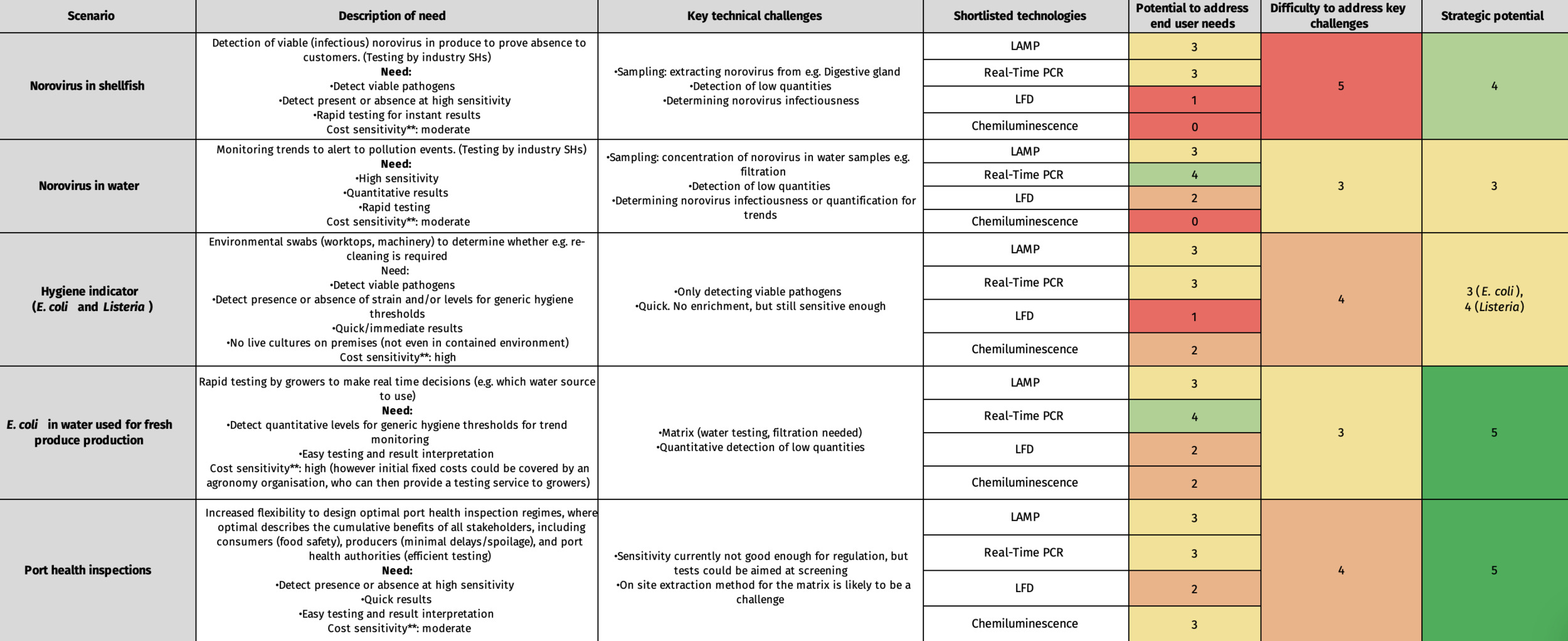
3.5. Pilot study
3.5.1. Portable real-time PCR for monitoring of E. coli in irrigation water.
3.5.1.1. Real-time PCR assay selection
To select a real-time PCR assay to use in the test, multiple assays from the literature and a commercial kit were compared for their sensitivity and specificity. The initial comparison of E. coli assay sensitivity showed that the Srinivasan et al. assay detected E. coli to the 10-4 dilution and therefore showed the lowest sensitivity (Appendix B). Silkie et al. detected both replicates down to the 10-6 dilution, however the R2 value dropped below 0.99 after the 10-5 dilution suggesting limit of quantification would be below this level. The Walker et al. assay could detect E. coli to the 10-5 dilution, however, the R2 value decreased after the 10-4 dilution. As the Silkie et al. assay showed the most promising sensitivity, it was taken forward for full comparison of analytical sensitivity and specificity with the commercial kit BioPoo® E. coli RT-PCR Go-Strips® (Biomeme).
The limit of detection of the Silkie et al. assay was 100 fg DNA, determined by the last dilution where all technical replicates from both DNA dilution series were detected as positive. The limit of detection of the E. coli commercial kit was 10 fg DNA. Both assays had R2 values over 0.99 down to the limit of detection (Figure 7). The assays start at very similar Ct values and then diverge due to difference in PCR efficiency. The Silkie et al. assay had 100% PCR efficiency, whereas the commercial kit had 156% PCR efficiency, with the Ct values increasing less than the standard 3.3 cycles per 10-fold dilution.
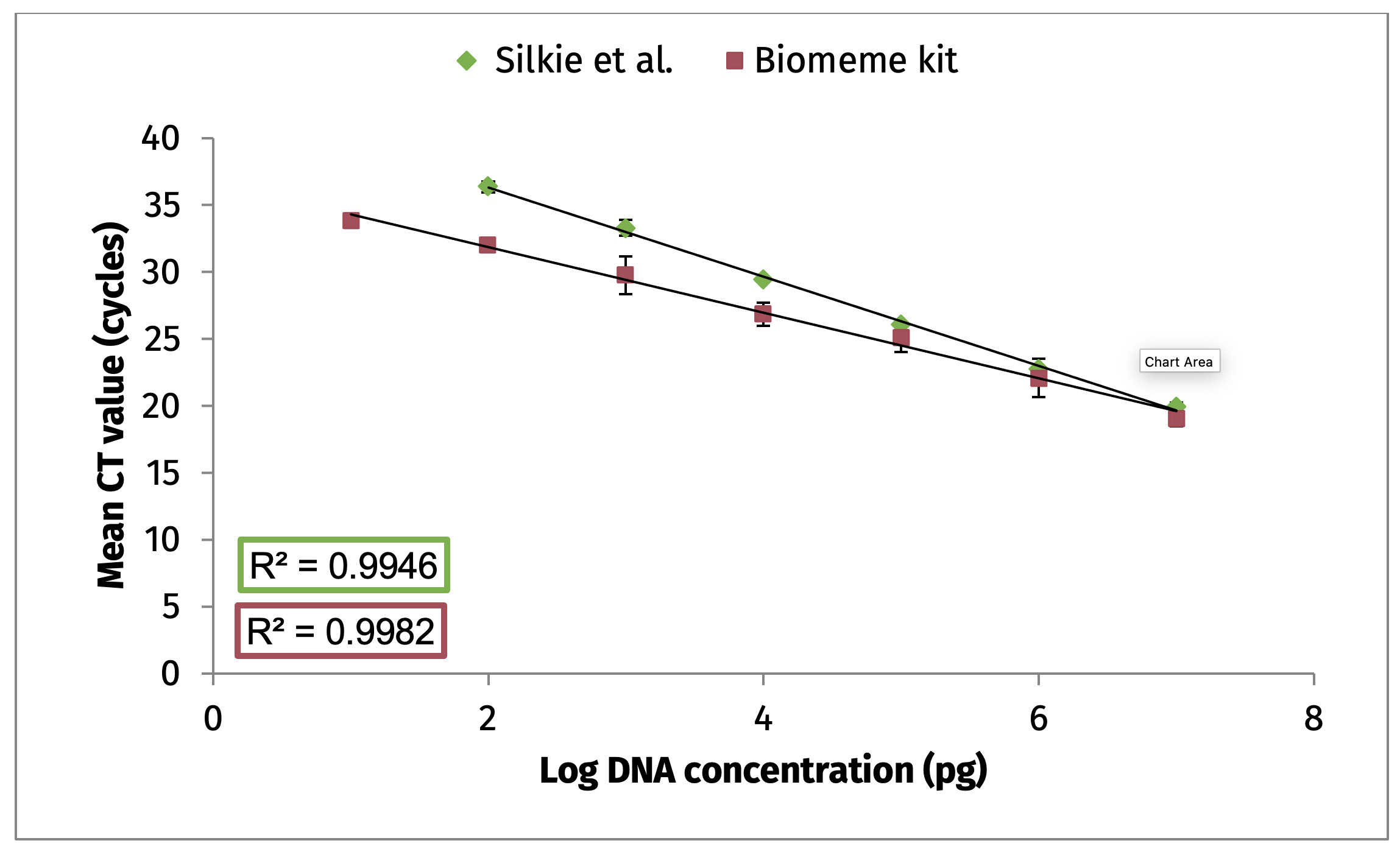
The Silkie et al. assay was tested on the Franklin thermocycler (Biomeme) to account for any difference which may be present due to the instrument and app software rather than the PCR assay. On the Franklin instrument the Silkie et al. assay showed very similar PCR efficiency of 101%, therefore the differences in PCR efficiency are due to differences in the assays themselves. The commercial kit is a multiplex assay and runs on much faster cycling conditions, so this might account for the difference. There were some differences identified when running on the Franklin instrument, which included lower Ct values and the 10 fg dilution being detected, so a potentially higher sensitivity is associated with the instrument. However, at the lower dilutions the Ct values standard deviations increased and the R2 value was reduced.
Isolates of target and non-target bacteria acquired from Fera’s culture collection and University of Lincoln were tested on the Silkie et al. assay and the commercial kit for detection of E. coli (Table 7). Both assays had 100% inclusivity of the E. coli isolates tested. Twenty-seven non-target isolates were tested for exclusivity, mostly consisting of closely related species which may cross react with the assays. The Silkie et al. assay had two potential cross reactions including Acinetobacter lwoffii, where the two isolate tested were giving late positive results (Ct >35), and Salmonella enterica where the isolate tested was giving inconsistent late positive results (Ct 39.76; Appendix B). No cross reactions were identified with the commercial kit. The results of the internal positive control (IPC) test in the commercial kit confirmed that all reactions were successful. Additional DNA extracts from isolates of E. coli isolated from river water provided CEFAS were used to improve the inclusivity panel and tested on the commercial kit only. Due to the slight advantages in terms of both analytical sensitivity and specificity and the major advantages in terms of ease of use for end-users the commercial kit BioPoo® E. coli RT-PCR Go-Strips® were chosen as the final assay for the pilot study.
3.5.1.2. Equipment free sample preparation and DNA extraction
Two methods of water filtration suitable for on-site deployment were trialled with water samples spiked with E. coli. Filtration method A1 produced lower Ct values (Table 12), suggesting that less E. coli cells are lost during this method, and it has higher sensitivity. A sample was also collected from the river Ouse (York) homogenised by mixing and processed by both methods and the gold standard to test the method practicality with a real sample containing typical inhibitors and particulate matter. The river sample had little E. coli within the sample, but the 16s assay seemed to suggest that the gold standard method was most effective at extracting bacterial DNA followed by on-site method A1 then A2 (data not shown). In terms of ease-of-use method A1 was also better, with fewer steps and easier filtration of turbid samples.
Filtration method A1 was chosen as the final method and used to compare an equipment free DNA extraction method using the M1 Sample Prep Cartridge DNA-HI (Biomeme), to the DNeasy PowerWater kit (Qiagen) as a gold standard method. Ct values increased by an average of ~1 (Table 12), when using the on-site method and therefore very little sensitivity was lost over the gold standard and the method was acceptable for the final test.
3.5.1.3. Assessment of test performance
The analytical sensitivity of the final method was assessed using a dilution series of E. coli pure culture The method reliably detected E. coli down to 103 CFU/100 ml, with 90% of samples detected at this level and therefore this is considered the limit of detection (LOD) (Table 13). Below this detection dropped off rapidly with only 17% of samples detected at 102 CFU/100 ml. The cell counts on plates of the LOD were calculated to be ~2000 CFU/ml. The Ct values were used to plot a standard curve which had an R2 value of >0.99 (Figure 8). Repeatability and reproducibility were assessed at a dilution close the limit of detection, both users detected 100% of samples and technical replicates (Appendix B). The average Ct value from all samples was very similar between the users 32.12 and 32.67 respectively showing good reproducibility.
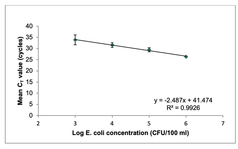
To determine the diagnostic performance of the test on real samples irrigation water sources were identified by end-users and samples collected and tested by the real-time PCR method and the gold standard method for enumeration of E. coli in water. All samples tested showed some reduction in fluorescence in IPC included in the commercial kit compared to clean water samples, however this decrease in fluorescence did not result in an increase in the Ct value. This could suggest that there is some inhibition in the real-time PCR using DNA extracts from real samples. The DNA extracts of two samples were spiked with E. coli DNA and tested, no increase in Ct value was seen compared to the clean E. coli DNA sample, suggesting no major inhibition. Samples 8, 11, 18 and 19 from initial tests, were re-tested diluted 1:10 and with the addition of BSA to the reaction (Table 14). When diluted the positive samples showed the expected increase in Ct value for the detection of E. coli and therefore diluting would reduce the sensitivity of the test. In samples 18 and 19 which were originally negative on the test, they were positive when diluted indicting that inhibition was affecting the original result. The IPC control Ct values stayed similar however the fluorescence values of the curves were improved. For samples 8 and 11 the addition of BSA had a small but positive effect on the fluorescence of the E. coli assay making the results graphs easier to assess and suggesting some inhibition is remediated. For samples 18 and 19, addition of BSA produced positive results. Therefore, the addition of BSA was recommended, and used in the final assessment of the real samples.
Out of the 25 samples tested, 5 samples were positive for E. coli on the portable real-time PCR test (Table 15). The positive samples included the samples with the highest E. coli counts according to the gold standard method, both the samples over >1000 CFU/ 100 ml were positive, and out of the nine samples between 100-1000 CFU/ 100 ml three samples were positive. Samples under 1000 CFU/100 ml would not be expected to be consistently picked up as they are under the LOD determined from previous experiments. The hygiene standard for irrigation water for high-risk crops is <100 CFU/100 ml sample, and therefore this criterion was used to calculate the diagnostic sensitivity, diagnostic specificity, positive predictive value, and negative predictive value of test compared to the gold standard. The diagnostic specificity of the assay was 100% as all true negative samples (<100 CFU/100 mL) were detected as negative by the method under validation, whereas diagnostic sensitivity was 45.45% as true positive samples (>100 CFU/100 mL) were missed by the real-time PCR method, likely due to the analytical sensitivity limitation described above. Positive and negative predictive values (PPV, NPV) indicate the likelihood that a sample which has a positive/negative test result does/does not have the pathogen being tested for and were 100% and 70% respectively.
Predictive values should only be interpretated if the number of positive and negative samples used for comparative testing approximates the ratio of positive and negative samples submitted for testing in a real scenario. The samples tested were from a range of real irrigation sources, however the time of year the comparison testing took place does not match the period real testing is likely to take place in summer, and therefore environmental factors may have influenced the samples collected.
Finally, the Ct values from the real-time PCR test did not show a relationship with CFU counts from the gold standard experiments. The standard curve generated in the sensitivity experiments would predict much higher E. coli levels from the Ct values from real samples tested (Figure 8). This could be due to levels non-viable E. coli DNA present in the sample which would contribute to the Ct value in the PCR but not to the CFU counts in the gold standard method. Therefore, the results from the real-time PCR would be difficult to relate to CFU values from the gold standard method and would need further work to give an indication of quantity. This lack of correlation may also reflect variable recovery efficiency during filtration and extraction, or matrix-related effects on amplification efficiency. Further work to integrate an internal process control in order to calculate the recovery efficiency may improve interpretation of the results.
3.5.1.4. Training with end-users, parallel testing, and feedback
Two end-users were trained to use the technology at Fera and then performed the method in a non-laboratory environment with real sample (Figure 9). User 1 processed three samples, and user 2 processed four samples and positive and negative controls were included in the testing. As part of parallel testing, duplicate water samples tested on-site were also tested in the laboratory using the portable real-time PCR method, and the DNA extracted by end-users on-site was also analysed in the laboratory to compare the PCR results, finally the water samples were also tested by the gold standard method. Overall, the DNA extractions performed by end users had 100% agreement with the samples processed in the laboratory (Table 16). However, the results from the real-time PCR performed on-site were often inconclusive with the IPC showing negative results meaning the reaction failed and should be repeated. The addition of the DNA extract to the real-time PCR reaction involves accurate pipetting, and the results indicate that this precision step is key and may require additional training.

3.5.2. LAMP for detection of Salmonella in sesame seeds at ports
3.5.2.1. LAMP assay selection
In the preliminary testing with dilutions of Salmonella Typhimurium smscoo1, amplification in negative controls was observed for assay 2, therefore this assay was not included in subsequent validation experiments. On the other hand, no non-specific amplification was detected for assays 1 and 3, and detection could be observed till 10-6 dilutions, therefore both assays were taken forward for further experiments (Appendix B).
A preliminary comparison of analytical sensitivity of the assay 1 and 3 was done using 10-fold dilutions of isolates Salmonella Enteritidis (024793_5/5) and Salmonella. sp. (023777_1/4). The limit of detection for both assays was determined to be 10-5 where positive results were obtained for both isolates. The performance of assays 1 and 3 was comparable as both assays showed the similar time to positive values across different dilutions and annealing temperatures were also consistent (Table 17).
Twenty Salmonella isolates and 25 non-target bacteria obtained from Fera’s culture collection, and the University of Lincoln were tested for assays 1 and 3 (Table 9). Both assays were able to detect all Salmonella isolates, showing 100% inclusivity and no cross-reaction with non-target bacterial isolates was observed. Additionally, to improve the specificity panel, 26 Salmonella isolates obtained from CEFAS were tested using assay 3 and all detected as positive by the assay.
Following the sensitivity and specificity experiments, equivalent results were obtained from LAMP assays 1 and 3. However, assay 3 was chosen for further experiments as it is commercially available in the form of a kit consisting only of master mix and primer mix therefore, is more user-friendly compared to assay 1, which requires handling master mix and six individual primers.
3.5.2.2. Development and testing of crude extraction method.
To develop the crude extraction methods, 103 CFU/ml was used for all experiments. The most basic approach was used as a start. Thus, in method B1, spiked seeds were shaken in 100 ml water and following heat lysis, 5 µl water was directly added to the LAMP reaction. Out of 5 samples tested, only one showed positive results (Table 18). It was anticipated that sensitivity would be compromised in this method due to the dilution effect caused by adding 100 ml of water. A smaller amount of water, 50 ml, was also attempted (data not shown), but this approach did not yield promising results.
To enhance the sensitivity, filtration was done using an analytical filter funnel (Nalgene™) equipped with a 0.45 µm pore filter paper in combination with a hand-operated vacuum pump (Nalgene™). In method B2, cells were dislodged from filter paper and heat lysed. Positive results were observed for white and brown seeds, while black seeds did not yield positive results. Since the extract from black seeds was coloured, it was hypothesised that the coloration compounds might be inhibiting the detection of LAMP results. Therefore, the DNA extracts from black seeds were diluted at a ratio of 1:5, and to further enhance the amplification, 0.5 µl of 50 mg/ml BSA was incorporated into the LAMP reaction. Positive results were observed following dilution and BSA, as illustrated in Table 19. The performance of BSA was also assessed with other varieties of seeds, and no alteration in the performance of assay or method was detected, therefore, the LAMP reaction was modified to add 0.5 µl BSA following 14.5 µl master mix and 5 µl primer mix.
In method B3, filtration followed by the Bento Dipstick DNA Extraction Kit yielded positive results for two out of five samples (Table 18). Meanwhile, method B4, involving filtration and the Qiagen Blood and Tissue Kit, demonstrated higher sensitivity for white and brown seeds and failed to yield positive results for black seeds. For this method, the addition of BSA and dilution did not affect the results.
Since Method B2 showed the most promising results, it was decided to proceed with this method, and the representation of all steps is shown in Figure 5.
3.5.2.3. Assessment of test performance
The analytical sensitivity of the final method was assessed using a dilution series of Salmonella sp. (023777_1/4). The method reliably detected Salmonella down to 103 CFU/25 g of seeds for all varieties tested therefore, this was considered the limit of detection (LOD; Table 20). The cell counts on the plate of LOD were calculated to be ~750 CFU/ml. No detection was observed below the dilution limit. At LOD, some variation in time to positive (Tp) values was observed across different seeds.
Results from heat-treated Salmonella broth for dilutions ranging from 105 to 101 showed that loss of sensitivity was not significant in the crude DNA extraction process as the LOD for heat-treated dilutions was similar to the overall method.
All biological and technical replicates tested by users 1 and 2 were successfully detected (Appendix B). The average time to positive for user 1 was 13:02 (±01:45) minutes and the annealing temperature was 87.95°C (±0.05°C). For user 2, the average detection time was 15:24 (±01:20) minutes and the annealing temperature was 88.13°C (±0.18°C) (Table 21).
To test the method on real samples, a total of 10 samples were received from Suffolk Coastal Port Health Authorities in Felixstowe over two months. It included a range of black, mixed, white, and brown seed varieties from countries such as Bolivia, India and China. Testing done by both UKHSA and Fera indicated that all samples were negative for Salmonella (Table 22). This may be attributed to the fact that the exporters are rigorously testing the samples before shipment therefore, the risk has been reduced in recent times.
3.5.2.4. Training with end-users, parallel testing and feedback
In the first stage of training at Fera, with a briefing of the method protocol and a demonstration of the method, the port health officials gained a theoretical and practical understanding of the overall method, quality control measures, result interpretation and troubleshooting. Subsequently, the end-users actively engaged in practicing the method themselves. By performing the method under guided supervision, the officials gained practical experience and skills in executing the procedure.

In the second stage, following on-site training, end-users independently tested the samples for one month (Figure 10). During this period, a total of 20 samples, comprising four shipments with five samples each, were tested by the end-users (Table 23). On-site testing, lab testing at Fera and UKHSA results indicated that all 5 samples within shipment 1, 3 and 4 were negative for Salmonella. Positive and negative control worked well in all LAMP assays, confirming the reliability and accuracy of the testing procedure.
In shipment 2, out of 5 seed samples tested, three showed positive detection on LAMP at the port and Fera. False positives through user contamination were ruled out by duplicate laboratory testing. The possibility of carryover contamination originating from the LAMP positive control or high-copy LAMP amplicons was considered however, it is less likely as training strongly emphasised disposal of LAMP strips immediately after amplification and that strips are never opened post-amplification. Also, the positive control DNA was added last after negative control and samples. All five samples were negative for Salmonella from gold standard testing at UKHSA. Shipment 2 DNA extracts from the port, when tested at Fera, showed positive detection in one out of five samples, compared to three out of five at the port. Given that crude extracts are not very stable, nucleic acid degradation might be speculated during shipment as it experienced some delays. Further gold standard testing by culturing method at Fera confirmed no Salmonella. Three bacteria were cultured from that sample: Acinetobacter baumannii, Pantoea sp., and Cronobacter malonaticus/turicensis. Their identity was confirmed morphologically and by sequencing 16S rRNA gene. Cross-reaction with these non-target organisms was ruled out by running LAMP on heat treated extracts of these bacteria that showed no amplification. There is potential that not all bacteria present on the sample were cultured during subsequent experiments. It could be hypothesised that non-viable Salmonella DNA present in the sample would be detected by the LAMP test but not the gold standard methods.
According to feedback from the end-users, training is crucial for the effective use of the technology and the method is easier and less-time consuming once the users are trained and experienced (Annex 5). At port, processing five samples from one shipment took approximately two hours. The accuracy of the results compared to the gold standard method was a key consideration. End-users raised concerns regarding the amount of plastic waste generated during the testing process. They agreed that the technology displayed promise for regular pathogen monitoring in informal samples and its suitability for formal or enforcement purposes remained uncertain. In the future, the technology could have potential applications in monitoring pathogens in ship food preparation areas and testing water quality. At ports, deployment of the technology would require infrastructure changes, including additional staff, regular trainings and equipment supplies. From the perspective of port health authorities, sending samples to UKHSA is more suitable as they receive allocations from them, incurring no additional time and equipment costs, and ensuring credible results. For the adoption of the technology, factors such as diagnostic performance, time to result, and cost ranked high in importance, followed by ease of use and sample throughput.
4. Discussion
To understand the landscape of rapid diagnostic technologies which could be deployed at the point of need we completed a wide scope literature review crosscutting multiple disciplines. It was clear in the review that efforts to develop diagnostic technologies for non-laboratory use increased in response to the COVID-19 pandemic. Development of point-of-care (POC) diagnostics for human health increasingly aspires towards complete integration of testing in stand-alone, preferably instrument-free, formats. From a technical perspective, many pathogen detection approaches described in this report, including those based on nucleic acid amplification, monoclonal antibodies and various biosensing modalities, have the potential to be incorporated into fully integrated devices combining a rapid sample-to-answer workflow and low per-test costs. However, these devices were not yet fully realised for markets focusing on food-borne pathogen detection.
The TRL framework was used to assess technologies in their maturity and closeness to deployment. The database (Annex 1) created with the technologies and their TRL for each in-scope pathogen provides a snapshot which can be used beyond this project to see which technologies are close to deployment in each area. The framework describes a roadmap of the next steps which would need to be undertaken to progress the technology towards deployment and could give an indication of effort and time to develop a technology. Following on from this study the TRL framework could be further expanded to provide detailed guidance and criteria which should be met before deploying an on-site technology in the food sector. The TRL framework is also transferrable outside of the project and could be used as a basis to assess diagnostic technologies in the future.
Most of the technologies assessed during the study did not progress beyond TRL 5 for in-scope pathogens (Figure 6), meaning that individual component technologies have been applied and validated for the detection of the in-scope pathogens, but have not been combined with other necessary components to form a sample-to-result test. For example, many of the nucleic acid-based detection technologies may have an amplification chemistry and detection method which could be feasibly performed on-site but have not demonstrated a suitable nucleic acid extraction method for the in-scope pathogen/matrix. Limited technologies achieved TRL 6 for the in-scope pathogens, where the performance of a component technology has been demonstrated in conjunction with other components necessary to form a sample-to-result solution. However, evidence of the technology’s performance in a relevant non-laboratory setting and in the hands of the intended users wasn’t present in the literature.
The lack of technologies progressing past TRLs 5/6 in our framework may be because academic funding sources through grants tend to focus on research and innovation at the early TRLs. Therefore, when a technology reaches a certain maturity, funding sources become limited, unless they are further developed and funded through the private sector. This funding gap between innovation and commercialisation is sometimes referred to as the ‘Valley of Death’ (Gbadegeshin et al., 2022). Research conducted by private companies may be less likely to be published in the scientific literature. Indeed, although we encountered commercially available kits aimed at the food-borne pathogen sector for some of the technologies, validation data in the hands of end-users in the deployment scenario was not readily available if it had taken place. Additionally, when diagnostic technologies progressed from academic research to private companies, they became much harder to find within the scope of the project.
However, it is likely most technologies are never developed past TRL 5 as many of the novel technologies such as biosensors, and microfluidic chips would not be feasible outside of an integrated device format. Development and commercialisation of integrated devices has historically been slow, taking many years from invention to deployment, and is extremely expensive, typically tens of millions of dollars (Berger et al., 2021; Chin et al., 2012). As a result, commercialisation of sample-to-answer diagnostic devices has been limited to only the highest priority clinical targets. More recent innovations in chip design and manufacture (e.g., 3D printing) may reduce both the time and costs associated with development of integrated diagnostic devices, but these barriers are likely to remain in the short term, especially for tests for which the market is small or unproven.
When shortlisting technologies for WP3 the focus was on technologies which could progress beyond TRL 5 to testing with end-users within the project. Therefore, we chose technologies which could work outside an integrated device format. While integration into a fully automated device with no manual steps may be an aspiration for any testing to be performed outside a laboratory, for some applications this may not be strictly necessary, for example, where testing will be carried out in relatively controlled conditions (e.g., a rudimentary laboratory), or where end-users could be provided with appropriate training and support to carry out more complex operations.
Development of an integrated device to a point where interaction with end-user was possible was unrealistic to reach within the scope of the project due to investment and time needed. Collaboration with, and feedback from end-users was rarely seen during our literature review, so a focus on end-user engagement to progress through the TRLs was prioritised. Working with end-users helps to identify problematic steps which may not have been recognised as such during development or laboratory validation. For some food testing applications, efforts focusing on end-user engagement, training and support, and in-field validation are more likely to lead to successful operational deployment of testing than technical innovations in miniaturisation and automation, for which the financial barriers to commercialisation may be too high.
Technologies chosen for the pilot study, real-time LAMP for testing of Salmonella in sesame seeds at ports and real-time PCR for detection of E. coli in irrigation water at farms, were at TRL 5. During the pilot study the technologies progressed to TRL 6 where a sample-to-result workflow suitable for on-site use in the targeted scenario was developed and validated. E. coli is a commonly used faecal indicator bacteria and is the recommend bacteria for testing fresh water sources (Allende et al., 2018), there is no legal requirement for testing of E. coli in irrigation water, however testing requirements are set by buyers such as supermarkets through assurance schemes. These schemes usually stipulate a limit of between ≤ 1000 CFU per 100 ml (Tesco Stores Nurture Scheme RTE irrigation water standards) or ≤ 100 CFU per 100 ml (Marks & Spencer Field to Fork RTE irrigation water standard) (Food Standards Scotland, 2024). For Salmonella there is a zero tolerance in a 25 g sample and a legal requirement for testing in many food products. Sesame seeds were chosen as the target matrix due to previous reports of Salmonella contamination in sesame seed products leading to the port health authorities regularly testing samples.
Both piloted technologies were DNA-based methods with high sensitivity. However, all commercially available laboratory-based kits using these technologies, particularly for Salmonella detection, required a pre-enrichment step to achieve the sensitivity required to meet regulations (Rani et al., 2021). Incorporating an enrichment step may be possible in rudimentary laboratories at the ports, but logical issues would have to be overcome, including appropriate facilities and training of end-users to ensure their safety when handling pathogens. The feasibility of this could be explored in a further study on successful deployment of on-site tests. Enrichment steps are also undesirable as they would reduce the rapidity of the tests. Neither piloted method included an enrichment step, and therefore their sensitivity did not equal the gold standard microbiology methods.
E. coli detection in irrigation water using portable real-time PCR had an analytical sensitivity of around 1000 CFU per 100 ml sample, and a diagnostic sensitivity of 46% based on the requirement to detect samples containing over 100 CFU E. coli. If a ten-fold increase in sensitivity could be achieved this technology could be a useful tool for detecting contaminated irrigation water. We were limited in our ability to optimise the real-time PCR as it came in the form of a commercial kit, if we explored outside this format increases in sensitivity may be achieved. The analytical and diagnostic specificity of the test was 100%. The ability to determine the general degree of contamination of water sources as well as noticing trends over time is highly desirable, however, the pilot study results did not indicate the method could provide quantitative information. The study was limited in the number of positive samples tested, as most fell below the limit of detection, if a much higher sample number were tested patterns may emerge. Previous research on quantitative PCR for E. coli water quality monitoring has demonstrated positive correlations with viable cells in river and freshwater bathing beaches, with 98% correct regulatory action (Lavender & Kinzelman, 2009). The integration of a non-target process control prior to filtration would allow recovery efficiency and inhibition to be assessed and allow. Results interpretation by end-users was a concern in the portable real-time PCR and associated software as results graphs could sometimes be ambiguous. The software also offers a summary of the results as positive or negative, but this could sometimes be incorrect and therefore interpretation of the graphs was critical.
Salmonella testing of sesame seeds via LAMP showed excellent analytical specificity, which is a key criterion for Salmonella testing due to many closely related species within Enterobacteriaceae. The analytical sensitivity of the test was around 1000 CFU in a 25 g sample. Although this does not meet current regulatory requirements, there may be a place for the test as a rapid screening tool for highly contaminated samples. Therefore, reducing the number of samples required for laboratory testing. On-site diagnostic tests could be considered as part of a testing programme, and if the trade-offs in performance are acceptable will depend on the conditions and operational context in which a test is performed (Buja et al., 2021) as well as the testing objective (Rainford et al., 2023).
A clear example of where an on-site method with a sensitivity much lower than the gold standard test has been successfully deployed is LFD as a screening tool for diagnosis of COVID-19. However, the use of LFDs would have had limited benefits if disease incidence was low, reinforcing the need to understand the operational context and objective of testing (Rainford et al., 2023). Whether a less sensitive but rapid on-site test would be a useful screening tool for Salmonella, would require further investigation. For the Salmonella testing in sesame seeds at ports, we were unable to calculate the diagnostic performance of the tests as all real samples received were negative for Salmonella. This is likely due to a recent industry procedural change to pre-test sesame seed products before export, and therefore only high-quality samples are now reaching the UK.
The test procedure for LAMP would benefit from the integration of an IPC to support reliable identification of false negative results. It will allow confirmation that the amplification process has functioned correctly and to distinguish true negative results from false negatives arising from inhibition, extraction failure, or reaction failure. This will be especially important for deployment with non-specialist end-users.
The pilot study also progressed the technologies within TRL 7 and both technologies were shown to be capable of being performed in the relevant environments with end-users and feedback was gathered. In the case of the pilot study of Salmonella detection at ports the end-users were able to independently perform the test over a one-month period and achieved the same results as duplicate samples performed by scientists in a laboratory setting using the same method. This is promising as the method involved multiple handling steps, accurate pipetting, or other precision operations which are vulnerable to contamination or misinterpretation of results. Therefore, could have been prone to a lack of reproducibility, generation of false results or high rates of retesting. During parallel testing there was one potential false positive result when compared to gold standard testing results. A full evaluation of diagnostic performance would be needed to see if this is a reoccurring issue of concern.
To reach TRL 8 a full evaluation of performance is required over a longer period of processing real samples until sufficient testing has been carried out to provide an explicit statistical understand of diagnostic performance in the relevant scenario. This information can then be used to consider potential outcomes of different deployment strategies. To reach full deployment of on-site testing technologies other logistical constraints will have to be addressed, and evidence requirements met to inform strategic/policy deployment decisions. This could include waste handling and decontamination procedures, reagent storage and traceability, end-user training requirements, and proficiency testing. Also, some level of physical separation between sample preparation areas and amplification or detection areas would be needed for sensitive tests. Further work is needed to clarify the requirements for integrating these technologies for the purpose of official controls in the food sector. One example could be aligning performance assessment with a recognised framework i.e. the ISO 16140 series.
Feedback on the technologies from end-user was important in revealing potential logistical barriers to deployment even if all technical requirements are met, including infrastructure change and legislative change requirements. A structured cost–benefit analysis of adoption comparing on-site testing versus submission to accredited laboratories (including cost, turnaround time, training burden, and regulatory acceptance) would provide a clearer basis for determining where these technologies offer genuine operational advantage. The development of official guidelines applicable across multiple types of on-site technologies could provide a framework for achieving deployment and increase the likelihood of realising on-site testing in the food sector.
Acknowledgements
This work is funded by the PATH-SAFE program from the HM Treasury Shared Outcome Funds. We would like to thank the Fera Science Ltd. core team for the contributions to the project: Emiline Quill, Jayne Hall, Jenny Tomlinson, Catherine Harrison, Barbara Agstner, and Rosario Romero, as well as our collaborator from the University of Lincoln, Bukola Onarinde.
We would also like to thank Fera Science Ltd. staff who contributed to the project: Abi Fisk, Joy Kaye, Christopher Field, Yue Lin Loh, Marco Benucci, Jen Clemens, Kinda Alraiss, Martin Boughtflower, Neil Taylor, Femke van den Berg, Nikolaos Tzanotis, Chris Conyers, Eleanor Jones, Valeria Orlando, Sarah Carroll, Lucy Brown.
We would also like to thank the end-uses who participated in the pilot study for their contribution to the project.
Special thanks to Edward Haynes and Rachel Baird, FSA for their invaluable help and support.